Review
. 2022 Nov 24:10:1046617.
doi: 10.3389/fcell.2022.1046617. eCollection 2022.

Mechanics and regulation of cytokinetic abscission

Affiliations
Review

Mechanics and regulation of cytokinetic abscission

Virginia Andrade et al. Front Cell Dev Biol. .

Abstract

Cytokinetic abscission leads to the physical cut of the intercellular bridge (ICB) connecting the daughter cells and concludes cell division. In different animal cells, it is well established that the ESCRT-III machinery is responsible for the constriction and scission of the ICB. Here, we review the mechanical context of abscission. We first summarize the evidence that the ICB is initially under high tension and explain why, paradoxically, this can inhibit abscission in epithelial cells by impacting on ESCRT-III assembly. We next detail the different mechanisms that have been recently identified to release ICB tension and trigger abscission. Finally, we discuss whether traction-induced mechanical cell rupture could represent an ancient alternative mechanism of abscission and suggest future research avenues to further understand the role of mechanics in regulating abscission.

Keywords: ESCRT; abscission; actin; caveolae; cell division; cell mechanics; cytokinesis; myosin II.


Conflict of interest statement

The authors declare that the research was conducted in the absence of any commercial or financial relationships that could be construed as a potential conflict of interest.

Figures

FIGURE 1
FIGURE 1
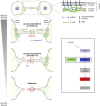
– Cytokinesis: A multi-step process. Step1 - Contraction of an actomyosin ring (orange) during furrow ingression. Step2 - Formation of the intercellular bridge (ICB) and midbody containing antiparallel microtubules (grey lines). Step3 - Thinning of the ICB. Step4 - Recruitment of the ESCRT-III to the midbody (MB). Step5 - Polymerization of ESCRT-III (red curves) on one side of the midbody and severing of the microtubules. Step6/7—Scission of the ICB membrane (abscission) and release of the midbody remnant (Step7). Adapted from (Andrade et al., 2022).
FIGURE 2
FIGURE 2
– Decrease in tension at the ICB triggers ESCRT-III polymerization and successful abscission in epithelial cells. Left panel: Decrease in the ICB tension and reduction of actomyosin contractility at the Entry Points (EPs) allow ESCRT-III polymerization, as cells progress towards abscission. Top right panel: Tension results from a combination of membrane tension and actomyosin-dependent cortical tension. Box: Schematic summarizing the contribution of the actomyosin network to the ICB tension, ESCRT-III polymerization and abscission. Y27632: drug that inhibits ROCK and prevents the phosphorylation (activation) of myosin II.
FIGURE 3
FIGURE 3
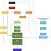
– Summary of the hierarchical events involved in the recruitment of the ESCRT-III machinery at the midbody and abscission site. MKLP1 at the midbody recruits both CEP55 and AKTIP. CEP55 directly recruits ALIX and TSG101, which act as two parallel pathways to recruit ESCRT-I/-II/-III at the midbody. ESCRT-III polymerizes to the abscission site (AS). Syntenin and Syndecan-4 stabilize ESCRT-III filaments at the AS. Orange: ESCRT-I related proteins; Light green: ESCRT-II components; Dark green: ESCRT-III components. Yellow: ALIX and associated complex (Syntenin, Syndecan-4). In cyan: Relationship between tension, the abscission checkpoint and ESCRT polymerization at the ICB. Only components of the abscission checkpoint likely involved in response to ICB tension have been depicted.
FIGURE 4
FIGURE 4
– Caveolae regulate ICB tension and ESCRT-III polymerization at the abscission site. A- Working model for caveolae-mediated regulation of the ICB tension. Note the progressive disappearance of caveolae (where asterisks are represented), possibly by flattening, at the Entry points while cells progress toward abscission. This membrane flattening is proposed to contribute to a decrease of the ICB tension, which promotes ESCRT-III polymerization and abscission. B- Depletion of caveolae leads to abscission delay, which can be corrected by either Y27632 (ROCK inhibitor) or hyperosmotic shock treatments. C- Schematic summary of events regulated by caveolae at the ICB. Adapted from (Andrade et al., 2022). EPs: Entry Points.

References

    1. Adar-Levor S., Nachmias D., Gal-Oz S. T., Jahn Y. M., Peyrieras N., Zaritsky A., et al. (2021). Cytokinetic abscission is part of the midblastula transition in early zebrafish embryogenesis. Proc. Natl. Acad. Sci. U. S. A. 118, e2021210118. 10.1073/pnas.2021210118 - DOI - PMC - PubMed
    1. Addi C., Bai J., Echard A. (2018). Actin, microtubule, septin and ESCRT filament remodeling during late steps of cytokinesis. Curr. Opin. Cell Biol. 50, 27–34. 10.1016/j.ceb.2018.01.007 - DOI - PubMed
    1. Addi C., Presle A., Frémont S., Cuvelier F., Rocancourt M., Milin F., et al. (2020). The Flemmingsome reveals an ESCRT-to-membrane coupling via ALIX/syntenin/syndecan-4 required for completion of cytokinesis. Nat. Commun. 11, 1941. 10.1038/s41467-020-15205-z - DOI - PMC - PubMed
    1. Agromayor M., Carlton J. G., Phelan J. P., Matthews D. R., Carlin L. M., Ameer-Beg S., et al. (2009). Essential role of hIST1 in cytokinesis. Mol. Biol. Cell 20, 1374–1387. 10.1091/mbc.e08-05-0474 - DOI - PMC - PubMed
    1. Amaral N., Vendrell A., Funaya C., Idrissi F. Z., Maier M., Kumar A., et al. (2016). The Aurora-B-dependent NoCut checkpoint prevents damage of anaphase bridges after DNA replication stress. Nat. Cell Biol. 18, 516–526. 10.1038/ncb3343 - DOI - PubMed

LinkOut - more resources