Review
. 2022 Dec 5:17:6007-6029.
doi: 10.2147/IJN.S386585. eCollection 2022.

A Comprehensive Review of the Application of Nanoparticles in Diabetic Wound Healing: Therapeutic Potential and Future Perspectives

Affiliations
Review

A Comprehensive Review of the Application of Nanoparticles in Diabetic Wound Healing: Therapeutic Potential and Future Perspectives

Wenqi Qin et al. Int J Nanomedicine. .

Abstract

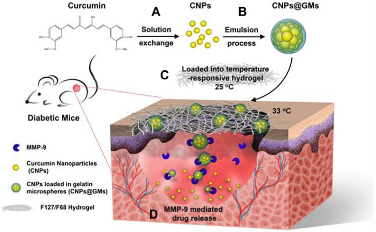
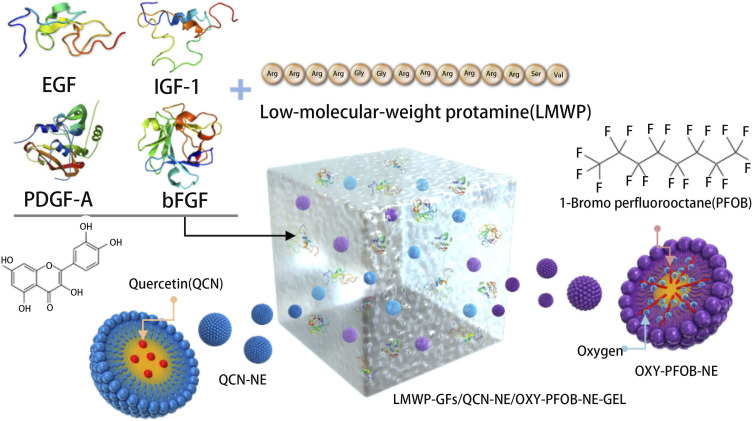
Diabetic wounds are one of the most challenging public health issues of the 21st century due to their inadequate vascular supply, bacterial infections, high levels of oxidative stress, and abnormalities in antioxidant defenses, whereas there is no effective treatment for diabetic wounds. Due to the distinct properties of nanoparticles, such as their small particle size, elevated cellular uptake, low cytotoxicity, antibacterial activity, good biocompatibility, and biodegradability. The application of nanoparticles has been widely used in the treatment of diabetic wound healing due to their superior anti-inflammatory, antibacterial, and antioxidant activities. These nanoparticles can also be loaded with various agents, such as organic molecules (eg, exosomes, small molecule compounds, etc.), inorganic molecules (metals, nonmetals, etc.), or complexed with various biomaterials, such as smart hydrogels (HG), chitosan (CS), and hyaluronic acid (HA), to augment their therapeutic potential in diabetic wounds. This paper reviews the therapeutic potential and future perspective of nanoparticles in the treatment of diabetic wounds. Together, nanoparticles represent a promising strategy in the treatment of diabetic wound healing. The future direction may be to develop novel nanoparticles with multiple effects that not only act in wound healing at all stages of diabetes but also provide a stable physiological environment throughout the wound-healing process.

Keywords: biomaterials; diabetic complications; diabetic wound; nanoparticles; wound healing.


Conflict of interest statement

The authors report no conflicts of interest in this work.

Figures

Figure 1
Figure 1
The healing process of diabetic wounds and normal wounds. Unlike normal wounds, diabetic wounds are characterized by hypoxia, impaired angiogenesis, chronic inflammation, and excessive expression of matrix metalloproteinases (MMPs), leading to the delayed healing of diabetic wounds.
Figure 2
Figure 2
Schematic illustration of the categories and therapeutic mechanism of biomaterials used on diabetic wounds. Biomaterials are loaded with organic molecules (including protein-loaded, exosomes, and small molecule compounds, etc.) and inorganic molecules (including AgNPs, CeNPs, SiO2NPs, etc.) to promote diabetic wound healing.
Figure 3
Figure 3
Schematic illustration showing the synthesis of F127-PEI, APu, multifunctional FEP scaffold dressing, nanoscale exosome-loaded FEP scaffold dressing and the potential application in diabetic wound healing and skin reconstruction.
Figure 4
Figure 4
Schematic representations of the CNPs@GMs/hydrogel preparation and the process of drug release at the wound bed in diabetic mice. (A) Preparation of pure CNPs via a solution exchange method. (B) CNPs loaded into GMs by an emulsion process to obtain CNPs@GMs. (C) The CNPs@GMs mixed with thermosensitive hydrogel and covered the wound in diabetic mice. (D) Under the microenvironment of a nonhealing wound, GMs were degraded by MMPs and specifically released the drug.
Figure 5
Figure 5
Schematic illustration of LMWP-GFs, QCN-NE, OXY-PFOB-NE, and a hydrogel comprising LMWP-GFs, QCN-NE, and OXY-PFOB-NE. Dove Medical Press Ltd.
Figure 6
Figure 6
Development of a transdermal drug delivery system for DFO and its regulation in the HIF-1a signaling pathway.
Figure 7
Figure 7
Diagrams displaying the formation of NIM-loaded micelles, VAN-AgNCs, hydrogels, and mechanisms of accelerating wound healing.

Similar articles

Cited by

References

    1. Cahill D, Zamboni F, Collins MN. Radiological advances in pancreatic islet transplantation. Acad Radiol. 2019;26(11):1536–1543. doi:10.1016/j.acra.2019.01.006 - DOI - PubMed
    1. Okonkwo UA, DiPietro LA. Diabetes and wound angiogenesis. Int J Mol Sci. 2017;18:7. doi:10.3390/ijms18071419 - DOI - PMC - PubMed
    1. Kesharwani P, Gorain B, Low SY, et al. Nanotechnology based approaches for anti-diabetic drugs delivery. Diabetes Res Clin Pract. 2018;136:52–77. doi:10.1016/j.diabres.2017.11.018 - DOI - PubMed
    1. Li SJ, Fan J, Zhou J, Ren YT, Shen C, Che GW. Diabetes mellitus and risk of bronchopleural fistula after pulmonary resections: a meta-analysis. Ann Thorac Surg. 2016;102(1):328–339. doi:10.1016/j.athoracsur.2016.01.013 - DOI - PubMed
    1. Shen YI, Cho H, Papa AE, et al. Engineered human vascularized constructs accelerate diabetic wound healing. Biomaterials. 2016;102:107–119. doi:10.1016/j.biomaterials.2016.06.009 - DOI - PubMed