. 2022 Nov 23:13:1052644.
doi: 10.3389/fphar.2022.1052644. eCollection 2022.

Postbiotic muramyl dipeptide alleviates colitis via activating autophagy in intestinal epithelial cells

Affiliations

Postbiotic muramyl dipeptide alleviates colitis via activating autophagy in intestinal epithelial cells

Yaying You et al. Front Pharmacol. .

Abstract

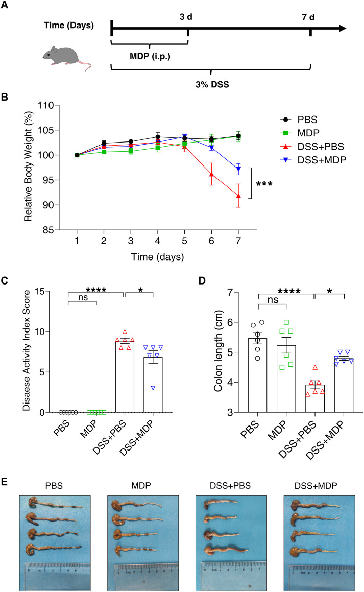
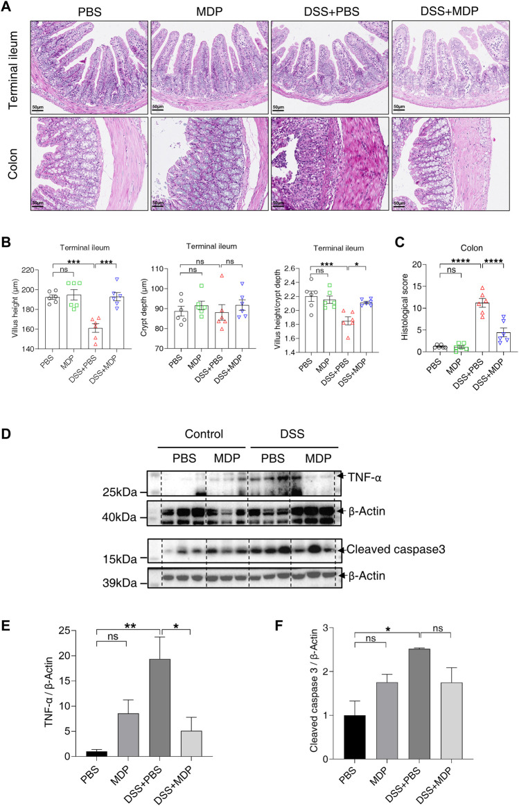
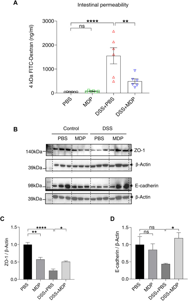
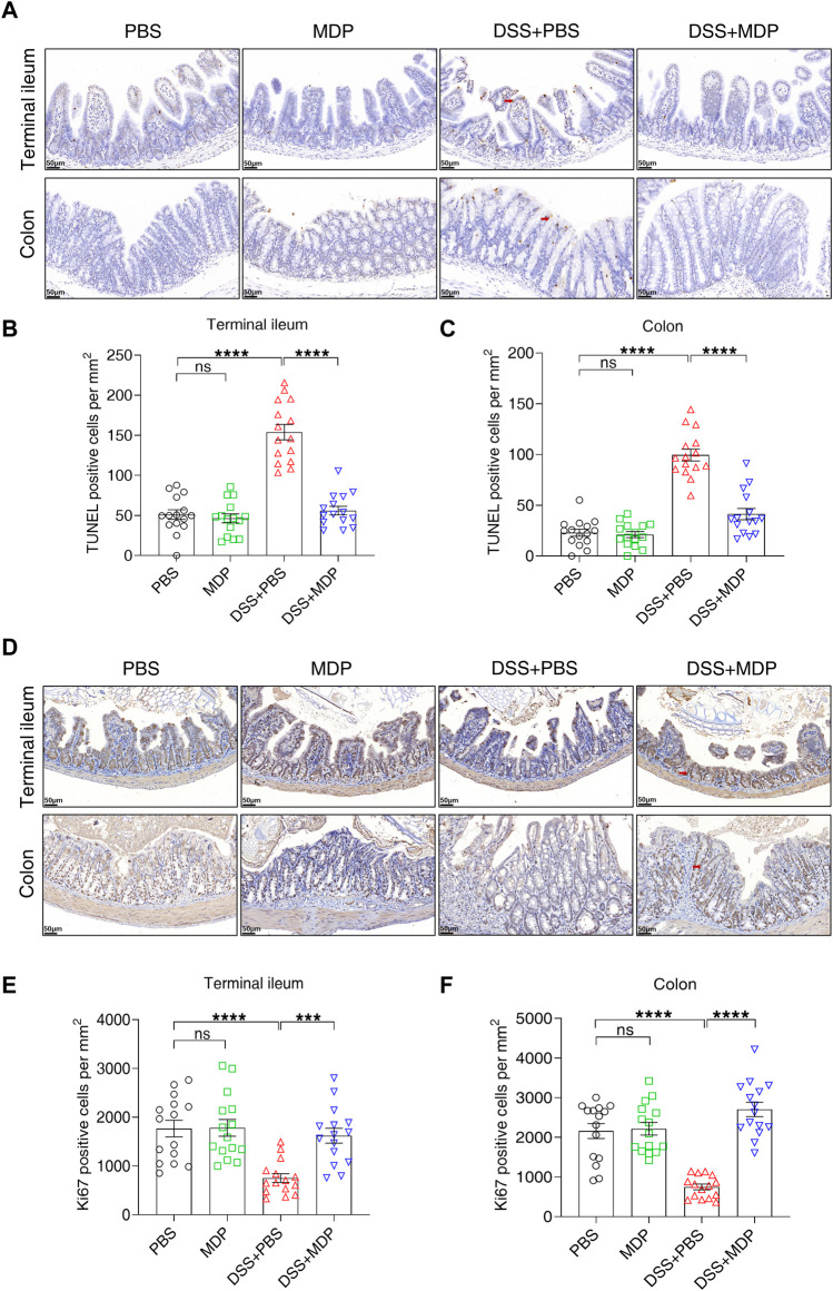
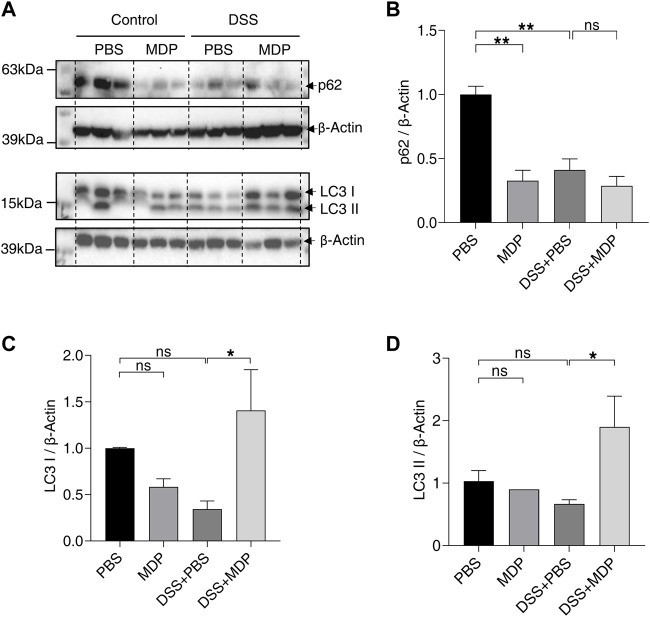
The pathogenesis of IBD is complicated and still unclear. Nucleotide-binding oligomerization domain 2 (NOD2) plays a significant role in regulating gut inflammation under the activation of muramyl dipeptide (MDP), which is used as a postbiotic. The study aimed to investigate the effect of MDP on the intestinal barrier in colitis and the mechanism involved. In this study, C57BL/6 mice were challenged with dextran sodium sulfate (DSS) for establishing a colitis model with the pre-treatment of MDP in vivo. Intestinal permeability was reflected by detecting the serum concentration of 4 kDa Fluorescein Isothiocyanate-Dextran. The expression of inflammation, barrier-related proteins, and autophagy was tested by Western Blotting. Proliferation and apoptosis in intestinal epithelial cells were detected by immunohistochemistry. Caco-2 cells were exposed to lipopolysaccharide for imitating inflammation in vitro. The findings showed that administration of MDP ameliorated losses of body weight loss, gross injury, and histology score of the colon in the DSS-induced colitis mice. MDP significantly ameliorated the condition of gut permeability, and promoted intestinal barrier repair by increasing the expression of Zonula occludens-1 and E-cadherin. Meanwhile, MDP promoted proliferation and reduced apoptosis of intestinal epithelial cells. In the experiment group treated with MDP, LC3 was upregulated, and p62 was downregulated, respectively. These results suggested that MDP stimulation attenuates intestinal inflammation both in vivo and in vitro. Potentially, MDP reduced the intestinal barrier damage by regulating autophagy in intestinal epithelial cells. Future trials investigating the effects of MDP-based postbiotics on IBD may be promising.

Keywords: MDP; NOD2; autophagy; colitis; intestinal barrier.


Conflict of interest statement

The authors declare that the research was conducted in the absence of any commercial or financial relationships that could be construed as a potential conflict of interest.

Figures

FIGURE 1
FIGURE 1
MDP alleviates DSS-induced intestinal injury in C57BL/6 mice. (A) Experimental timeline was shown. C57BL/6 mice were divided into four groups: PBS group, MDP group, DSS + PBS group, and DSS + MDP group (n = 6, each group). Each group received continuous intraperitoneal injections (i.p.) of MDP (100 μg) or sterile PBS 100 μL from Day 1 to Day 3, respectively. The mice drank water or 3% DSS from Day 1 to Day 7. (B) Body weight of mice was recorded every day. (C) Weight loss, stool consistency, and blood of mice were observed for evaluating disease activity index scores. (D,E) Mice were euthanized on Day 8. The colon was taken and the length was measured and photographed. Data were expressed as mean ± SEM. *p <0.05, ***p <0.001, ****p <0.0001, ns, not significant, One-way or Two-way ANOVA.
FIGURE 2
FIGURE 2
MDP inhibits inflammatory response in the intestine. (A) Representative images of H&E staining for terminal ileum and colon tissues in PBS, MDP, DSS + PBS, and DSS + MDP group mice (n = 6, each group), respectively. scale bars: 50 μm. (B) Quantification of villus height, crypt depth, and the ratio of villus height and crypt depth in the terminal ileum (n = 6, each group). (C) The histological score of colons (n = 6, each group). (D) Western blotting analysis for TNF-α and Cleaved caspase 3 with β-Actin as the internal standard protein in colon of PBS, MDP, DSS + PBS, and DSS + MDP group mice. Representative images of three duplicate samples of the immune blotting were shown. (E,F) Quantification of the relative expression of TNF-α and Cleaved caspase 3 in panel (D) by ImageJ, respectively. Data were expressed as mean ± SEM. *p <0.05, **p <0.01, ***p <0.001, ****p <0.0001, ns, not significant, One-way ANOVA.
FIGURE 3
FIGURE 3
MDP reduces DSS-induced intestinal barrier damage. (A) Mice were fasted overnight on Day 7 and given FITC-Dextran by gavage on Day 8. After 4 h, blood was taken from the eyeball from light. The serum was separated and analyzed for the detection of intestinal permeability (n = 6, each group). (B) Western blotting analysis for ZO-1 and E-cadherin with β-Actin as the internal standard protein in the colon of PBS, MDP, DSS + PBS, and DSS + MDP group mice. Representative images of three duplicate samples of the immune blotting were shown. (C,D) Quantification of the relative expression of ZO-1 and E-cadherin in panel (B) by ImageJ, respectively. Data were expressed as mean ± SEM. *p <0.05, **p <0.01, ****p <0.0001, ns, not significant, One-way ANOVA.
FIGURE 4
FIGURE 4
MDP decreases the apoptosis of intestinal epithelium in DSS-induced mice. (A) Representative images of TUNEL staining for terminal ileum and colon tissues in PBS, MDP, DSS + PBS, and DSS + MDP group mice (n = 3, each group), respectively. scale bars: 50 μm. Red arrows indicated positive cells. (B,C) Quantification of TUNEL positive cells per mm2 in terminal ileum and colon (n = 3, each group), respectively. (D) Representative images of Ki67 staining for terminal ileum and colon tissues in PBS, MDP, DSS + PBS, and DSS + MDP group mice (n = 3, each group), respectively. scale bars: 50 μm. Red arrows indicated positive cells. (E,F) Quantification of Ki67 positive cells per mm2 in terminal ileum and colon (n = 3, each group), respectively. Data were expressed as mean ± SEM. ***p <0.001, ****p <0.0001, ns, not significant, One-way ANOVA.
FIGURE 5
FIGURE 5
MDP activates autophagy in colon of DSS-induced mice. (A) Western blotting analysis for p62, LC3 І, and LC3 ІІ with β-Actin as the internal standard protein in the colon of PBS, MDP, DSS + PBS, and DSS + MDP group mice. Representative images of three duplicate samples of the immune blotting were shown. (B–D) Quantification of the relative expression of p62, LC3 І and LC3 ІІ in panel (A) by ImageJ, respectively. Data were expressed as mean ± SEM. *p <0.05, **p <0.01, ns, not significant, One-way ANOVA.
FIGURE 6
FIGURE 6
MDP reduces intestinal barrier damage and activates autophagy in vitro. (A) Schematic diagram of the administration of MDP (0, 1, 5, 10 μg/ml, respectively) in Caco-2 cells. (B) Western blotting analysis for NOD2, LC3 І, and LC3 ІІ with GAPDH as the internal standard protein in Caco-2 cells. Representative images of the immune blotting were shown. (C,D) Quantification of the relative expression of NOD2 and LC3 ІІ in panel (B) by ImageJ, respectively. Results were from three independent experiments. (E) Cytotoxicity of Caco-2 cells with the administration of with LPS (0, 1, 10, 100 μg/mL, respectively) for 24 h by testing LDH OD value. (F) Relative RNA levels of IL-1β of Caco-2 cells with the administration of LPS. (G) Western blotting analysis for ZO-1, p62, and LC3 ІІ with GAPDH as the internal standard protein in Caco-2 cells of the groups (Control, MDP, LPS, and LPS + MDP), respectively. Representative images of three duplicate samples of the immune blotting were shown. (H–J) Quantification of the relative expression of ZO-1, p62, and LC3 ІІ in panel (G) by ImageJ, respectively. (K) Cytotoxicity of Caco-2 cells of the groups (Control, MDP, LPS, and LPS + MDP) by testing LDH OD value. Data were expressed as mean ± SEM. *p <0.05, **p <0.01, ns, not significant, One-way ANOVA.

Similar articles

Cited by

References

    1. Aardoom M. A., Veereman G., de Ridder L. (2019). A Review on the use of anti-TNF in children and adolescents with inflammatory bowel disease. Int. J. Mol. Sci. 20 (10), E2529. 10.3390/ijms20102529 - DOI - PMC - PubMed
    1. Adak A., Khan M. R. (2019). An insight into gut microbiota and its functionalities. Cell. Mol. Life Sci. 76 (3), 473–493. 10.1007/s00018-018-2943-4 - DOI - PMC - PubMed
    1. Al Nabhani Z., Dietrich G., Hugot J. P., Barreau F. (2017). Nod2: The intestinal gate keeper. PLoS Pathog. 13 (3), e1006177. 10.1371/journal.ppat.1006177 - DOI - PMC - PubMed
    1. Alnabhani Z., Montcuquet N., Biaggini K., Dussaillant M., Roy M., Ogier-Denis E., et al. (2015). Pseudomonas fluorescens alters the intestinal barrier function by modulating IL-1β expression through hematopoietic NOD2 signaling. Inflamm. Bowel Dis. 21 (3), 543–555. 10.1097/mib.0000000000000291 - DOI - PubMed
    1. Arab H. H., Al-Shorbagy M. Y., Saad M. A. (2021). Activation of autophagy and suppression of apoptosis by dapagliflozin attenuates experimental inflammatory bowel disease in rats: Targeting AMPK/mTOR, HMGB1/RAGE and Nrf2/HO-1 pathways. Chem. Biol. Interact. 335, 109368. 10.1016/j.cbi.2021.109368 - DOI - PubMed

LinkOut - more resources