. 2022 Nov 24:13:1052091.
doi: 10.3389/fphar.2022.1052091. eCollection 2022.

Evaluation and manipulation of tissue and cellular distribution of cardiac progenitor cell-derived extracellular vesicles

Affiliations

Evaluation and manipulation of tissue and cellular distribution of cardiac progenitor cell-derived extracellular vesicles

Marieke T Roefs et al. Front Pharmacol. .

Abstract

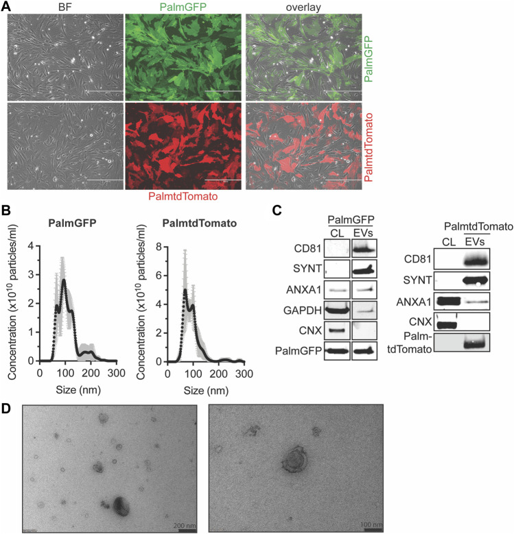
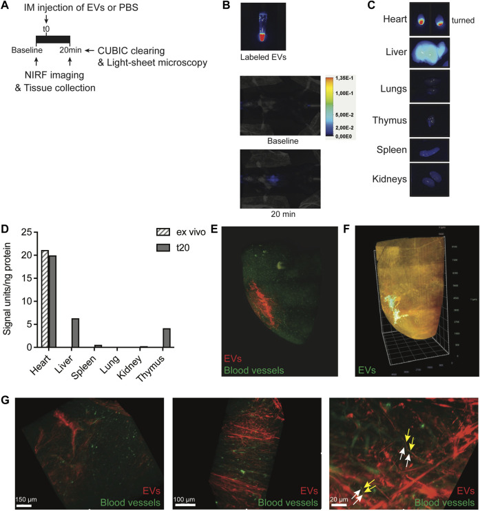
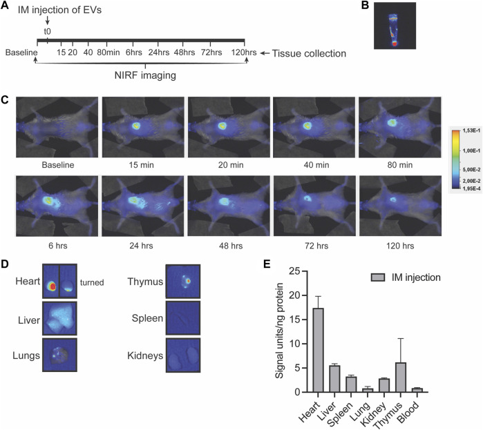
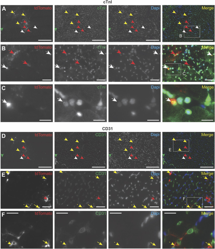
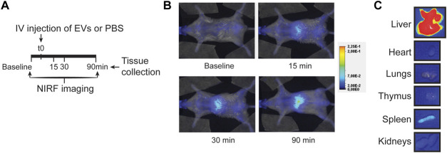
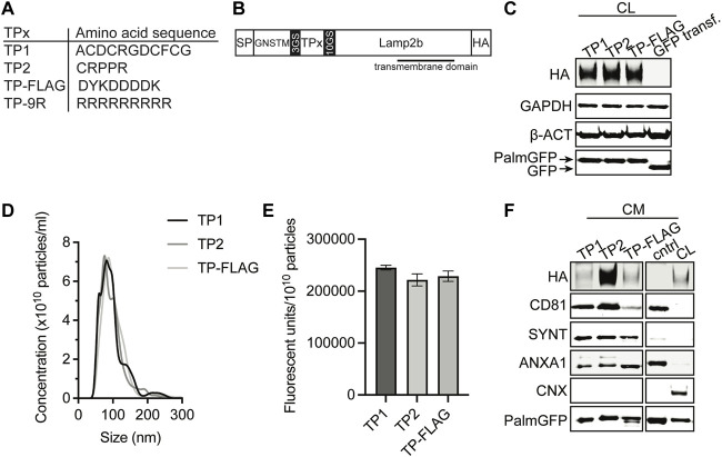
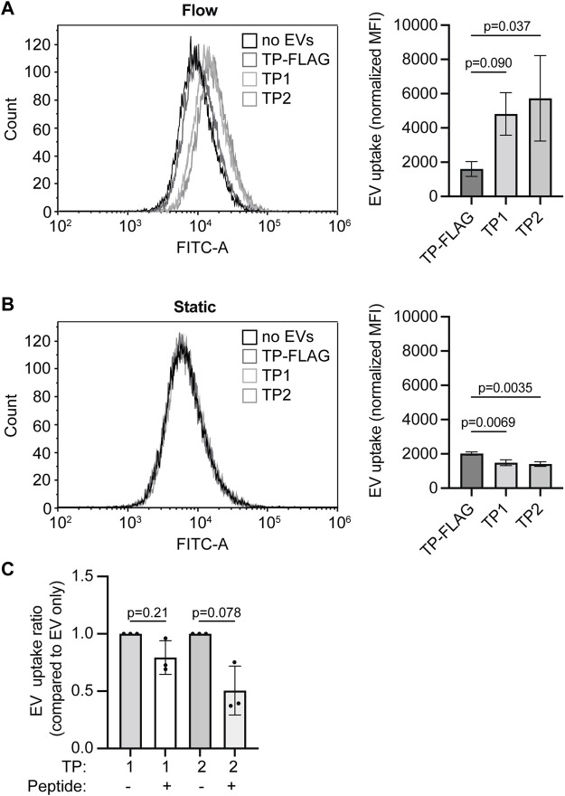
Cardiac progenitor cell-derived extracellular vesicles (CPC-EVs) have been successfully applied via different delivery routes for treating post-myocardial infarction injury in several preclinical models. Hence, understanding the in vivo fate of CPC-EVs after systemic or local, i.e. myocardial, delivery is of utmost importance for the further therapeutic application of CPC-EVs in cardiac repair. Here, we studied the tissue- and cell distribution and retention of CPC-EVs after intramyocardial and intravenous injection in mice by employing different EV labeling and imaging techniques. In contrast to progenitor cells, CPC-EVs demonstrated no immediate flush-out from the heart upon intramyocardial injection and displayed limited distribution to other organs over time, as determined by near-infrared imaging in living animals. By employing CUBIC tissue clearing and light-sheet fluorescent microscopy, we observed CPC-EV migration in the interstitial space of the myocardium shortly after EV injection. Moreover, we demonstrated co-localization with cTnI and CD31-positive cells, suggesting their interaction with various cell types present in the heart. On the contrary, after intravenous injection, most EVs accumulated in the liver. To potentiate such a potential systemic cardiac delivery route, targeting the cardiac endothelium could provide openings for directed CPC-EV therapy. We therefore evaluated whether decorating EVs with targeting peptides (TPs) RGD-4C or CRPPR connected to Lamp2b could enhance EV delivery to endothelial cells. Expression of both TPs enhanced CPC-EV uptake under in vitro continuous flow, but did not affect uptake under static cell culture conditions. Together, these data demonstrate that the route of administration influences CPC-EV biodistribution pattern and suggest that specific TPs could be used to target CPC-EVs to the cardiac endothelium. These insights might lead to a better application of CPC-EV therapeutics in the heart.

Keywords: biodistribution; endothelial cell; exosome; extracellular vesicle; heart; targeting.


Conflict of interest statement

The authors declare that the research was conducted in the absence of any commercial or financial relationships that could be construed as a potential conflict of interest.

Figures

FIGURE 1
FIGURE 1
Characterization of PalmGFP- and PalmtdTomato-labeled EVs derived from CPCs. (A) Fluorescent microscopy pictures of CPC cells stably expressing (top) PalmGFP or (bottom) PalmtdTomato. Scale bars represent 400 µm. (B) Representative NTA plots showing the size distribution and particle concentration of EVs after SEC isolation of conditioned medium derived from PalmGFP+- and PalmtdTomato+ CPCs. (C) Western blot analysis showing the presence of CD81, Syntenin-1 (SYNT), AnnexinA1 (ANXA1), GAPDH, PalmGFP and/or PalmtdTomato, and absence of Calnexin (CNX) in (left) PalmGFP+- and (right) PalmtdTomato+ EVs. Cell lysates (CL) derived from (left) PalmGFP+ or (right) (TdTomato-negative) CPCs were included as control. Uncut blots are included in Supplementary Figure S10. (D) Representative TEM images of PalmGFP+ EVs at two different magnifications.
FIGURE 2
FIGURE 2
Short-term EV biodistribution after intramyocardial injection. (A) Schematic overview of EV administration and near-infrared (NIRF) imaging directly or at 20 min after intramyocardial injection (IM) of 5 μL EVs in the left ventricle. (B) NIRF images taken from the Eppendorf containing labeled EVs and living animal at baseline and 20 min after intramyocardial EV administration. (C) NIRF images of individual organs collected after termination. Uncut images are included in Supplementary Figure S11. (D) Quantification of fluorescence (800 nm) per ng protein in organ lysates as compared to organ background. An ex vivo heart injected with 5 μL EVs was included as control. (E,F) Snap-shots of 3D fluorescent images of the heart injected with (E) 2.5 × 10¹⁰ or (F) 1.75 × 10¹⁰ AlexaFluor647 NHS ester-labeled EVs, generated after CUBIC tissue clearing and subsequent light-sheet fluorescent microscopy. Images represent two independent experiments. (E) Heart was perfused with Lectin-FITC to stain blood vessels before tissue collection. Snapshots at higher magnification are displayed in (G). Arrows indicate no co-localization of (yellow) EVs and (white) blood vessels.
FIGURE 3
FIGURE 3
Long-term EV distribution in the mouse after intramyocardial injection. (A) Schematic overview of intramyocardial (IM) EV administration in the left ventricle wall and near-infrared (NIRF) imaging up to 5 days after intramyocardial injection. (B,C) Representative NIRF images taken from (B) the eppendorf containing labeled EVs, (C) the living animal at various time points after EV administration and (D) of individual organs collected after 5 days follow-up. Uncut images are included in Supplementary Figure S11. (E) Quantification of fluorescence (800nm) per ng protein in organ lysates as compared to organ background. Data of two mice are shown and are displayed as mean ± SD.
FIGURE 4
FIGURE 4
Immunocytochemistry analysis of EV uptake in the heart. PalmtdTomato-EVs were administered in the left ventricle wall of a healthy mouse heart through intramyocardial injection and heart tissue was collected after 4 h (A–F) Immunofluorescence staining of two subsequent heart sections, cut across the sagittal plane, using antibodies against tdTomato (shown in red), and co-staining with antibodies against (A–C) cardiomyocyte marker cardiac Troponin I (cTnI, shown in green) and (D–F) blood vessel-specific CD31 (shown in green). (B) Enlargement of the square in panel A. (C) Enlargement of the square in panel B. (E) Enlargement of the square in panel D. (F) Enlargement of the square in panel E. Nuclei are visualized with DAPI (shown in blue). tdTomato co-localization with other stainings are indicated with arrows: cTnI (white), CD31 (yellow), no co-localization (red). Scale bars = 50 μm (A,D), 250 μm (B,E), 100 μm (C,F).
FIGURE 5
FIGURE 5
EV distribution in the mouse after intravenous injection. (A) Schematic overview of EV administration and NIRF imaging up to 90 min after tail vein injection. (B,C) Representative NIRF images taken (B) from the living animal at various time points after EV administration and (C) of individual organs collected after 90 min follow-up. Uncut images are included in Supplementary Figure S11.
FIGURE 6
FIGURE 6
Generation of EVs expressing targeting peptides for endothelial cells. (A) Amino acid sequence of TP1, TP2, TP-FLAG, which was included as negative control, and TP-9R. (B) Schematic of constructs expressed on EVs consisting of a specific targeting peptide (TPx) connected to transmembrane protein Lamp2b, flanked by a N-terminal signal peptide (SP) and GNSTM glycosylation sequence, and C-terminal HA-tag. (C) Representative western blot analysis showing expression of HA-tag and PalmGFP in cell lysate (CL) harvested from CPC lines stably expressing PalmGFP+ and specific TPs. CPCs transduced with GFP were included as negative control. GAPDH and beta-actin (ß-ACT) were included as house-keeping proteins. (D) Representative NTA plots showing the size distribution and particle concentration of concentrated conditioned medium (CM) derived from PalmGFP+ TP-expressing CPCs. (E) GFP fluorescence per 1010 particles determined in CM derived from PalmGFP+ TP-expressing CPCs (n = 2). Data are displayed as mean ± SD. (F) Representative western blot analysis showing expression of HA-tag, PalmGFP, CD81, Syntenin-1 (SYNT) and AnnexinA1 (ANXA1) in CM. CL of PalmGFP+ CPCs stably expressing TP-FLAG were included as control. CM derived from CPCs transduced with GFP was included as negative control (cntrl). Calnexin (CNX) was only present in CL. Uncut blots are included in Supplementary Figure S10.
FIGURE 7
FIGURE 7
Targeting peptide expression on EVs increases EV uptake in HMEC-1 under flow conditions. (A, B) EV-TP1, -TP2, and -TP-FLAG uptake in HMEC-1 under (A) flow or (B) static conditions, determined by flow cytometry. Mean fluorescence is corrected for negative control (M199 medium administration). (C) EV-TP1 and EV-TP2 uptake in HMEC-1 under flow after HMEC-1 pre-incubation of CRGDC or CRPPR peptides. For each replicate, mean fluorescence is corrected for the average of negative control, and conditions with peptides are compared to conditions without peptide pre-incubation. Data are displayed as mean ± SD and represent three replicate experiments.

Similar articles

Cited by

References

    1. Algoet M., Janssens S., Himmelreich U., Gsell W., Pusovnik M., Van den Eynde J., et al. (2022). Myocardial ischemia-reperfusion injury and the influence of inflammation. Trends cardiovasc. Med. 22, S1050–1738. 10.1016/J.TCM.2022.02.005 - DOI - PubMed
    1. Alvarez-Erviti L., Seow Y., Yin H., Betts C., Lakhal S., Wood M. J. A. (2011). Delivery of siRNA to the mouse brain by systemic injection of targeted exosomes. Nat. Biotechnol. 29, 341–345. 10.1038/nbt.1807 - DOI - PubMed
    1. Arslan F., Lai R. C., Smeets M. B., Akeroyd L., Choo A., Aguor E. N. E. E., et al. (2013). Mesenchymal stem cell-derived exosomes increase ATP levels, decrease oxidative stress and activate PI3K/Akt pathway to enhance myocardial viability and prevent adverse remodeling after myocardial ischemia/reperfusion injury. Stem Cell Res. 10, 301–312. 10.1016/j.scr.2013.01.002 - DOI - PubMed
    1. Avraamides C. J., Garmy-Susini B., Varner J. A. (2008). Integrins in angiogenesis and lymphangiogenesis. Nat. Rev. Cancer 8, 604–617. 10.1038/NRC2353 - DOI - PMC - PubMed
    1. Barile L., Cervio E., Lionetti V., Milano G., Ciullo A., Biemmi V., et al. (2018). Cardioprotection by cardiac progenitor cell-secreted exosomes: role of pregnancy-associated plasma protein-A. Cardiovasc. Res. 114, 992–1005. 10.1093/cvr/cvy055 - DOI - PubMed