

Modern imaging techniques for monitoring patients with multiple myeloma
- PMID: 36506611
- PMCID: PMC9694753
- DOI: 10.15386/mpr-2215
Modern imaging techniques for monitoring patients with multiple myeloma
Abstract
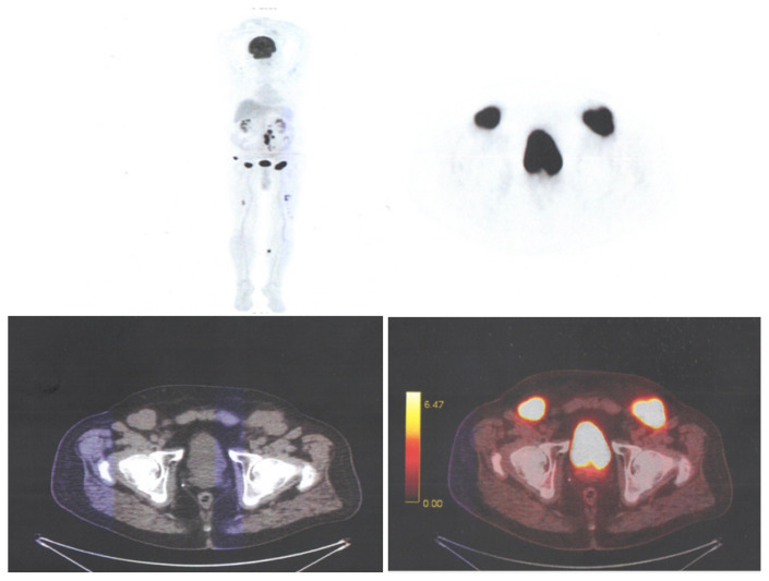
Bone disease is a serious problem for many patients, often causing pathological bone fractures. A spinal collapse is a condition that affects the quality of life. It is the most frequent feature of multiple myeloma (MM), used in establishing the diagnosis and the need to start treatment. Because of these complications, imaging plays a vital role in the diagnosis and workup of myeloma patients. For many years, conventional radiography has been considered the gold standard for detecting bone lesions. The main reasons are the wide availability, low cost, the relatively low radiation dose and the ability of this imaging method to cover the entire bone system. Because of its incapacity to evaluate the response to therapy, more sophisticated techniques such as whole-body low-dose computed tomography (WBLDCT), whole-body magnetic resonance imaging, and 18F-fluorodeoxyglucose-positron emission tomography/computed tomography (PET/CT) are used. In this review, some of the advantages, indications and applications of the three techniques in managing patients with MM will be discussed. The European Myeloma Network guidelines have recommended WBLDCT as the imaging modality of choice for the initial assessment of MM-related lytic bone lesions. Magnetic resonance imaging is the gold-standard imaging modality for the detection of bone marrow involvement. One of the modern imaging methods and PET/CT can provide valuable prognostic data and is the preferred technique for assessing response to therapy.
Keywords: 18F-fluorodeoxyglucose positron emission tomography-computed tomography (FDG-PET/CT); MRI; multiple myeloma; skeletal survey; whole-body low-dose computed tomography (WBLDCT).
Figures


Similar articles
-
Imaging in multiple myeloma: How? When?Blood. 2019 Feb 14;133(7):644-651. doi: 10.1182/blood-2018-08-825356. Epub 2018 Dec 26. Blood. 2019. PMID: 30587527 Review.
-
Skeletal Survey in Multiple Myeloma: Role of Imaging.Curr Med Imaging. 2021;17(8):956-965. doi: 10.2174/1573405617666210126155129. Curr Med Imaging. 2021. PMID: 33573573 Review.
-
Whole-body low-dose computed tomography and advanced imaging techniques for multiple myeloma bone disease.Clin Cancer Res. 2014 Dec 1;20(23):5888-97. doi: 10.1158/1078-0432.CCR-14-1692. Epub 2014 Oct 7. Clin Cancer Res. 2014. PMID: 25294899 Review.
-
More advantages in detecting bone and soft tissue metastases from prostate cancer using 18F-PSMA PET/CT.Hell J Nucl Med. 2019 Jan-Apr;22(1):6-9. doi: 10.1967/s002449910952. Epub 2019 Mar 7. Hell J Nucl Med. 2019. PMID: 30843003
-
A prospective comparison of 18F-fluorodeoxyglucose positron emission tomography-computed tomography, magnetic resonance imaging and whole-body planar radiographs in the assessment of bone disease in newly diagnosed multiple myeloma.Haematologica. 2007 Jan;92(1):50-5. doi: 10.3324/haematol.10554. Haematologica. 2007. PMID: 17229635
Cited by
-
The numerous facets of 1q21+ in multiple myeloma: Pathogenesis, clinicopathological features, prognosis and clinical progress (Review).Oncol Lett. 2024 Apr 9;27(6):258. doi: 10.3892/ol.2024.14391. eCollection 2024 Jun. Oncol Lett. 2024. PMID: 38646497 Free PMC article. Review.
References
-
- Leydon P, O’Connell M, Greene D, Curran K. Automatic bone marrow segmentation for PETCT imaging in multiple myeloma. Phys Med. 2016;32:242. - PubMed
-
- Colucci PG, Schweitzer AD, Saab J, Lavi E, Chazen JL. Imaging findings of spinal brown tumors: a rare but important cause of pathologic fracture and spinal cord compression. Clin Imaging. 2016;40:865–869. - PubMed
-
- Gouliamos A, Andreou J, Kosmidis P. Imaging in Clinical Oncology. doi: 10.1007/s00259-014-2875-7. - DOI
Publication types
LinkOut - more resources
Full Text Sources