

Loss of dental tissue after restoration or sealing of occlusal carious lesions: 3-4-year results of randomized clinical trials
- PMID: 36506636
- PMCID: PMC9733557
- DOI: 10.4103/jcd.jcd_194_22
Loss of dental tissue after restoration or sealing of occlusal carious lesions: 3-4-year results of randomized clinical trials
Abstract
Context: Although preservation of the tooth structure is quoted as the main advantage of sealing of carious lesions, there are no long-term studies comparing the maintenance of dental tissue after restoration or after caries sealing.
Aim: To measure the radiographically visible loss of dental tissue after conventional restoration and sealing of carious lesions.
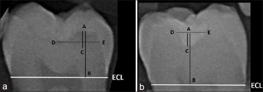
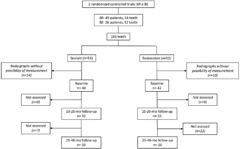
Subjects and methods: This study was a secondary analysis of two randomized controlled clinical trials, one conducted in Brazil and another in Belgium, which evaluated two different therapies for the treatment of occlusal carious lesions in permanent teeth: sealant (SE) without previous carious tissue removal or restoration (RE) with total removal of carious dentin. The greater depth and width of sealed carious lesions and restorations were compared.
Statistical analysis: The independent t-test was used to compare therapies at different time points, while the paired t-test was used to compare the same therapy over time.
Results: Carious lesions in the RE and SE groups showed similar measurements at baseline (P > 0.05). Over time, significantly greater loss of tooth structure was observed in the RE group than in the SE group. No increase in lesion depth or width was observed in the SE group, suggesting no progression of sealed lesions.
Conclusion: Sealing of carious lesions resulted in greater preservation of dental tissue.
Keywords: Dental caries; dental radiography; dental restoration; dental sealant; randomized clinical trial.
Copyright: © 2022 Journal of Conservative Dentistry.
Conflict of interest statement
There are no conflicts of interest.
Figures
Similar articles
-
Sealing of cavitated occlusal carious lesions in the dentine of deciduous molars: a two-year randomised controlled clinical trial.Clin Oral Investig. 2022 Jan;26(1):1017-1024. doi: 10.1007/s00784-021-04085-2. Epub 2021 Jul 21. Clin Oral Investig. 2022. PMID: 34286398 Clinical Trial.
-
A randomized clinical trial on the sealing of occlusal carious lesions: 3-4-year results.Braz Oral Res. 2017 Jun 5;31:e44. doi: 10.1590/1807-3107BOR-2017.vol31.0044. Braz Oral Res. 2017. PMID: 28591240 Clinical Trial.
-
Conventional caries removal and sealed caries in permanent teeth: a microbiological evaluation.J Dent. 2012 Sep;40(9):776-82. doi: 10.1016/j.jdent.2012.05.011. Epub 2012 Jun 2. J Dent. 2012. PMID: 22664566
-
Managing Carious Lesions: Consensus Recommendations on Carious Tissue Removal.Adv Dent Res. 2016 May;28(2):58-67. doi: 10.1177/0022034516639271. Adv Dent Res. 2016. PMID: 27099358
-
Contemporary concepts in carious tissue removal: A review.J Esthet Restor Dent. 2017 Nov 12;29(6):403-408. doi: 10.1111/jerd.12338. Epub 2017 Sep 19. J Esthet Restor Dent. 2017. PMID: 28925550 Review.
References
-
- Elderton RJ. Preventive (evidence-based) approach to quality general dental care. Med Princ Pract. 2003;12(Suppl 1):12–21. - PubMed
-
- Qvist V. Longevity of restorations: 'The death spiral'. In: Fejerskov O, Nyvad B, Kidd E, editors. Dental Caries: The Disease and Its Clinical Management. Hoboken: Wiley-Blackwell; 2005. pp. 387–401.
-
- Handelman SL, Washburn F, Wopperer P. Two-year report of sealant effect on bacteria in dental caries. J Am Dent Assoc. 1976;93:967–70. - PubMed
-
- Theilade E, Fejerskov O, Migasena K, Prachyabrued W. Effect of fissure sealing on the microflora in occlusal fissures on human teeth. Arch Oral Biology. 1977;22:251–9. - PubMed
-
- Oong EM, Griffin SO, Kohn WG, Gooch BF, Caufield PW. The effect of dental sealants on bacteria levels in caries lesions: A review of the evidence. J Am Dent Assoc. 2008;139:271–8. - PubMed
LinkOut - more resources
Full Text Sources