Case Reports
. 2022 Sep;23(3):327-335.
doi: 10.30476/DENTJODS.2021.90454.1491.

Multifocal Osteoblastoma of the Jaws: A Very Rare Case Report

Affiliations
Case Reports

Multifocal Osteoblastoma of the Jaws: A Very Rare Case Report

Mohammad Moshref et al. J Dent (Shiraz). 2022 Sep.

Abstract

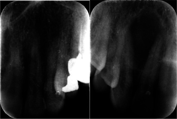
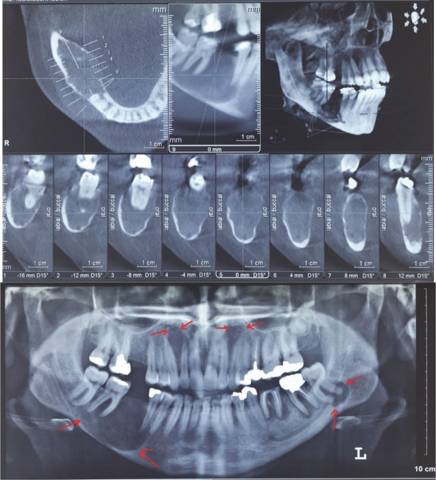
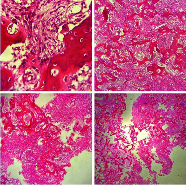
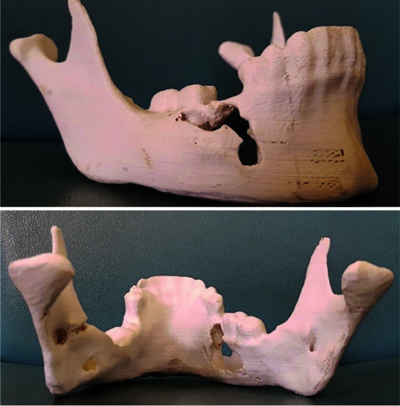
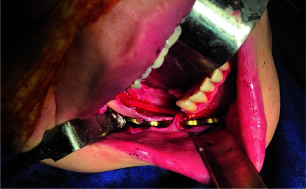
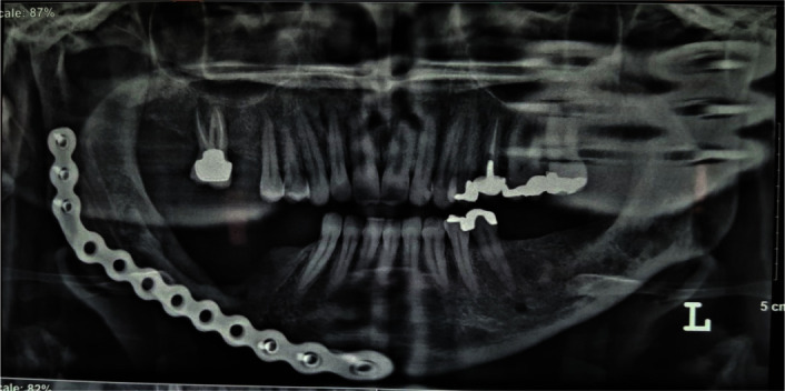
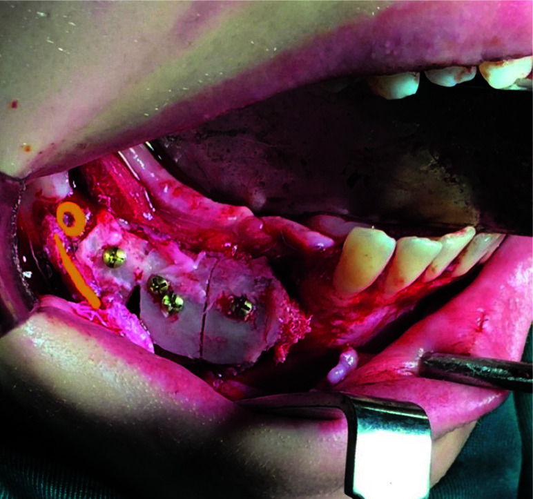
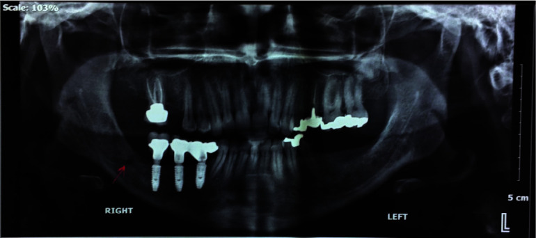
Osteoblastoma is a solitary benign bone-forming neoplasm, which comprises 1% of all primary bone tumors. Multifocal benign osteoblastoma of the jaws is very rare. Osteoblastoma must be differentiated from other similar bone-forming lesions such as osteoid osteoma and osteosarcoma for correct diagnosis and proper treatment planning. Therefore, precise examination of the patient and correlation with radiographic and histological features are essential for the best treatment and prognosis. This study reports a rare case of multifocal osteoblastoma in a 30-year-old female, involving the mandible and the maxilla, which was treated by surgical excision, iliac bone graft reconstruction, and implantation. Complete surgical excision is necessary to treat osteoblastoma with a good prognosis. The patient was followed-up for four years postoperatively, and there were no signs of recurrence in the panoramic view or the clinical examination.

Keywords: Enucleation; Jaws; Multifocal; Osteoblastoma; Peripheral ostectomy.


Conflict of interest statement

The authors declare that they have no conflict of interest.

Figures

Figure 1
Figure 1
Pre-operative panoramic view, arrows show multifocal lesion. Periapical view of maxilla lesion
Figure 2
Figure 2
Pre-operative cone-beam computed tomography (CBCT) view, buccal and lingual bone thinning, and perforation, push the inferior alveolar nerve toward the lower border
Figure 3
Figure 3
Histopathological images showed stroma containing osteoblasts, scattered dilated vascular channels, and large sheets of irregular trabeculae in osteoid material rimmed by osteoblast with ample cytoplasm and hyperchromatic nuclei
Figure 4
Figure 4
Pre-operative medical stereolithography (3D printing model of the mandible)
Figure 5
Figure 5
Intra-operative view, pre-bent reconstruction plate was fixed intraorally; the inferior alveolar nerve was preserved
Figure 6
Figure 6
Post-operative panoramic view, 6 months after operation
Figure 7
Figure 7
Cone-beam computed tomography (CBCT) after reconstruction with iliac bone graft
Figure 8
Figure 8
Intra-operative view (secondary surgery), iliac bone graft, the line shows inferior alveolar nerve and the circle shows new mental foramen
Figure 9
Figure 9
Post-operative panoramic view, reconstruction of the right mandible with implantation, arrow shows the new mental foramen position

References

    1. Cekic B, Toslak IE, Yildirim S, Uyar R. Osteoblastoma originating from frontoethmoidal sinus causing personality disorders and superior gaze palsy. Niger J Clin Pract. 2016;19:153–155. - PubMed
    1. Mardaleishvili K, Kakabadze Z, Machavariani A, Grdzelidze T, Kakabadze A, Sukhitashvili N, et al. Benign osteoblastoma of the mandible in a 12-year-old female: A case report. Oncol Lett. 2014;8:2691–2694. - PMC - PubMed
    1. Vella O, Cuny F, Robard L, Bazille C. Osteoblastoma of the maxillary sinus in a child presenting with exophthalmos. Eur Ann Otorhinolaryngol Head Neck Dis. 2016;133:277–279. - PubMed
    1. Mahajan A, Kumar P, Desai K, Kaul RP. Osteoblastoma in the retromolar region- Report of an unusual case and Review of literature. J Maxillofac Oral Surg. 2013;12:338–340. - PMC - PubMed
    1. Shah S, Kim JE, Huh KH, Yi WJ, Heo MS, Lee SS. Recurrent osteoblastoma of the maxilla. Dentomaxillofac Radiol. 2013;42:20100263. - PMC - PubMed

Publication types

LinkOut - more resources