

MRI Methods for Imaging Beta-Cell Function in the Rodent Pancreas
- PMID: 36507988
- PMCID: PMC10008468
- DOI: 10.1007/978-1-0716-2807-2_7
MRI Methods for Imaging Beta-Cell Function in the Rodent Pancreas
Abstract
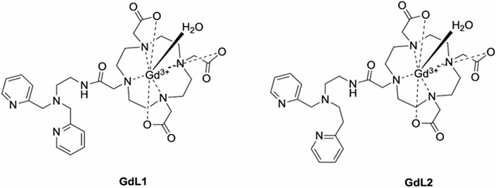
The role of Zn2+ ions in proper storage of insulin in β-cell granules is well-established so when insulin is secreted from β-cells stimulated by an increase in plasma glucose, free Zn2+ ions are also released. This local increase in Zn2+ can be detected in the pancreas of rodents in real time by the use of a zinc-responsive MR contrast agent. This method offers the opportunity to monitor β-cell function longitudinally in live rodents. The methods used in our lab are fully described in this short report and some MR images of a rat pancreas showing clearly enhanced hot spots in the tail are presented.
Keywords: Functional imaging; Glucose-stimulated insulin secretion (GSIS); Glucose-stimulated zinc secretion (GSZS); Imaging β-cell function in vivo; MRI; Rodent models; Zinc-responsive agents.
© 2023. The Author(s), under exclusive license to Springer Science+Business Media, LLC, part of Springer Nature.
Figures


Similar articles
-
Imaging Insulin Secretion from Mouse Pancreas by MRI Is Improved by Use of a Zinc-Responsive MRI Sensor with Lower Affinity for Zn2+ Ions.J Am Chem Soc. 2018 Dec 19;140(50):17456-17464. doi: 10.1021/jacs.8b07607. Epub 2018 Dec 11. J Am Chem Soc. 2018. PMID: 30484648 Free PMC article.
-
Imaging β-Cell Function Using a Zinc-Responsive MRI Contrast Agent May Identify First Responder Islets.Front Endocrinol (Lausanne). 2022 Jan 31;12:809867. doi: 10.3389/fendo.2021.809867. eCollection 2021. Front Endocrinol (Lausanne). 2022. PMID: 35173681 Free PMC article.
-
Autocrine effect of Zn²⁺ on the glucose-stimulated insulin secretion.Endocrine. 2015 Sep;50(1):110-22. doi: 10.1007/s12020-015-0568-z. Epub 2015 Mar 15. Endocrine. 2015. PMID: 25771886
-
Emerging molecular technologies for light-mediated modulation of pancreatic beta-cell function.Mol Metab. 2022 Oct;64:101552. doi: 10.1016/j.molmet.2022.101552. Epub 2022 Jul 19. Mol Metab. 2022. PMID: 35863638 Free PMC article. Review.
-
Transcription factors involved in glucose-stimulated insulin secretion of pancreatic beta cells.Biochem Biophys Res Commun. 2009 Jul 10;384(4):401-4. doi: 10.1016/j.bbrc.2009.04.135. Epub 2009 May 3. Biochem Biophys Res Commun. 2009. PMID: 19410555 Review.
Cited by
-
Neurodevelopmental Consequences of Dietary Zinc Deficiency: A Status Report.Biol Trace Elem Res. 2023 Dec;201(12):5616-5639. doi: 10.1007/s12011-023-03630-2. Epub 2023 Mar 25. Biol Trace Elem Res. 2023. PMID: 36964812 Review.
References
-
- Kim EE (2014) Clinical perfusion MRI: techniques and applications. J Nucl Med 55:522
-
- Hecht S, Adams WH (2010) MRI of brain disease in veterinary patients part 2: acquired brain disorders. Vet Clin North Am Small Anim Pract 240:39–63 - PubMed
-
- Kurniawan ND (2018) MRI in the study of animal models of neurodegenerative diseases. In: García Martín ML, López Larrubia P (eds) Preclinical MRI: methods and protocols. Springer, New York, pp 347–375 - PubMed
Publication types
MeSH terms
Substances
Grants and funding
LinkOut - more resources
Full Text Sources
Miscellaneous
