

Use of a Novel Three-dimensional Head-mounted Digital Visualization Platform in Corneal Endothelial Transplantation
- PMID: 36508107
- PMCID: PMC9834461
- DOI: 10.1007/s40123-022-00624-6
Use of a Novel Three-dimensional Head-mounted Digital Visualization Platform in Corneal Endothelial Transplantation
Abstract
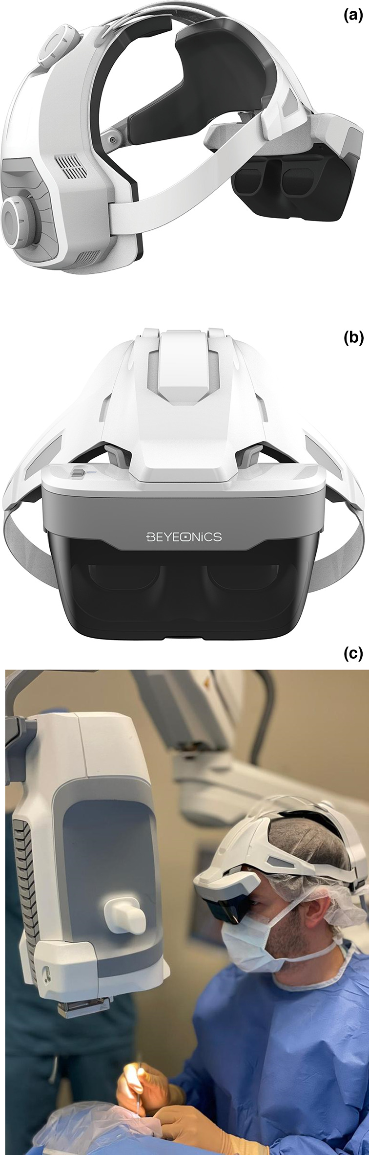
Introduction: To report the first endothelial keratoplasty procedures performed using a 3D digital head-mounted ophthalmic exoscope.
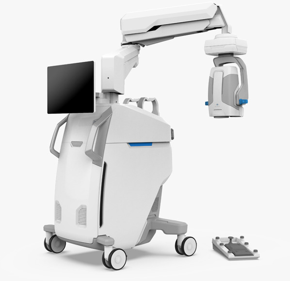
Methods: Three patients underwent Descemet stripping automated endothelial keratoplasty (DSAEK) using a 3D digital ophthalmic exoscope (Beyeonics One, Beyeonics Vision, Haifa, Israel) at the Tel Aviv Sourasky Medical Center, Tel Aviv, Israel.
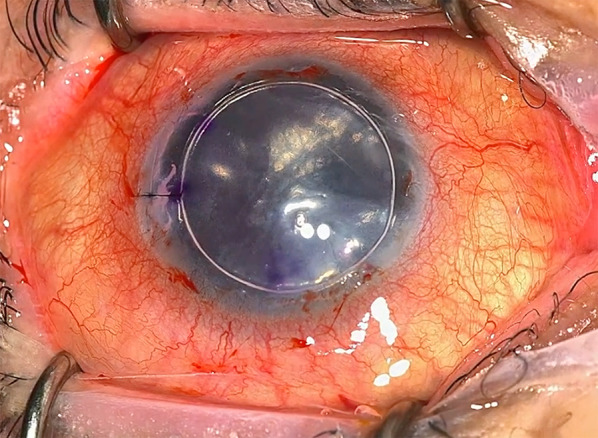
Results: All procedures were uneventful, leading to resolution of corneal edema and vision improvement. Surgeons reported excellent visualization and minimal lag, almost negligible, with the benefits of improved ergonomics and the use of head gestures to control zoom, focus, brightness, and panning. There were no postoperative complications.
Conclusion: The new 3D digital ophthalmic exoscope system can be successfully used in DSAEK surgery with potential advantages in ergonomics, picture quality, and image control. Further studies can compare this system with either standard operating microscopes or 3D heads-up display systems.
Keywords: Corneal endothelial transplantation; Descemet stripping automated endothelial keratoplasty (DSAEK); Exoscope; Head gesture; Head-mounted display (HMD); Heads-up; Microscope; Surgery; Three-dimensional (3D); Visualization.
© 2022. The Author(s).
Figures
References
-
- Keeler R. The evolution of the ophthalmic surgical microscope. Hist Ophthal Intern. 2015;1:35–66.
LinkOut - more resources
Full Text Sources
