. 2022 Dec 12;17(12):e0278058.
doi: 10.1371/journal.pone.0278058. eCollection 2022.

A Drosophila model of HPV16-induced cancer reveals conserved disease mechanism

Affiliations

A Drosophila model of HPV16-induced cancer reveals conserved disease mechanism

Lydia Hashemi et al. PLoS One. .

Abstract

High-risk human papillomaviruses (HR-HPVs) cause almost all cervical cancers and a significant number of vaginal, vulvar, penile, anal, and oropharyngeal cancers. HPV16 and 18 are the most prevalent types among HR-HPVs and together cause more than 70% of all cervical cancers. Low vaccination rate and lack of molecularly-targeted therapeutics for primary therapy have led to a slow reduction in cervical cancer incidence and high mortality rate. Hence, creating new models of HPV-induced cancer that can facilitate understanding of the disease mechanism and identification of key cellular targets of HPV oncogenes are important for development of new interventions. Here in this study, we used the tissue-specific expression technique, Gal4-UAS, to establish the first Drosophila model of HPV16-induced cancer. Using this technique, we expressed HPV16 oncogenes E5, E6, E7 and the human E3 ligase (hUBE3A) specifically in the epithelia of Drosophila eye, which allows simple phenotype scoring without affecting the viability of the organism. We found that, as in human cells, hUBE3A is essential for cellular abnormalities caused by HPV16 oncogenes in flies. Several proteins targeted for degradation by HPV16 oncoproteins in human cells were also reduced in the Drosophila epithelial cells. Cell polarity and adhesion were compromised, resulting in impaired epithelial integrity. Cells did not differentiate to the specific cell types of ommatidia, but instead were transformed into neuron-like cells. These cells extended axon-like structures to connect to each other and exhibited malignant behavior, migrating away to distant sites. Our findings suggest that given the high conservation of genes and signaling pathways between humans and flies, the Drosophila model of HPV16- induced cancer could serve as an excellent model for understanding the disease mechanism and discovery of novel molecularly-targeted therapeutics.


Conflict of interest statement

The authors have declared that no competing interests exist.

Figures

Fig 1
Fig 1. Expression of HPV16 E5, E6, and E7 oncogenes leads to mortality in a human UBE3A-dependent manner.
Transgenes were expressed using the GMR-Gal4 in the eye discs (A-D). The wild type (A) and expression of HPV16 E5, E6, E7 alone (B) or hUBE3A (C) showed no phenotypic defects in the adult fly eye. Flies co-expressing E5, E6, E7 and hUBE3A (D) did not develop beyond the pupal stage.
Fig 2
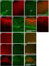
Fig 2. HPV16 E5, E6, E7 oncoproteins disrupt epithelial integrity.
Transgenes were expressed in the third instar larval eye discs (A-E’) and in pupal eyes 42 hours after pupation (H-P), using GMR-Gal4. Tissues were immunolabelled for adherens junction protein, E-cad and the expression of mCD8RFP was used to mark the area of transgenes expression. (A) GMR-Gal4-expressing eye disc showing an intact epithelium with proper localization of E-cad (A’) and a sequential pattern of differentiated ommatidia. (B-D) Co-expression of HPV16 E5, E6, E7 and the membrane marker, mCD8RFP showing no cellular abnormalities as indicated by proper localization of E-cad and ommatidia formation (C, C’). (E-G) Eye disc co-expressing HPV16 E5, E6, E7+hUBE3A+ mCD8RFP exhibited mislocalization or loss of E-cad (F, F’), rounded cell morphology (E, E’), and disrupted pattern of ommatidial formation (F). (H-J) Pupal eye 42 hrs after pupation expressing GMR-Gal4 and mCD8RFP showing a fully differentiated eye with a stereotype pattern of hexagonal array of ommatidia and proper localization of E-cad (I). (K-M) Pupal eye co-expressing HPV16 E5, E6, E7+ mCD8RFP showed no cellular abnormalities and eyes exhibited similar morphology and E-cad localization to the control eyes in H-J. (N-P) Pupal eye co-expressing HPV16 E5, E6, E7+hUBE3A+ mCD8RFP showed no ommatidia formation and instead the pupal eye was clusters of cells (N) that had lost cell-cell adhesion evident by lack of E-cad. (O). Insets are digitally magnified 200%. Scale bar indicates 100 μm in all panels except insets in which scale bar represents 50 μm.
Fig 3
Fig 3. Expression of HPV16 E5, E6, E7 oncogenes in conjunction with hUBE3A induces apoptosis in larval eye disc.
Transgenes were expressed in the third instar larval eye discs (A-I) and in pupal eyes 42 hours after pupation (J-R), using GMR-Gal4. mCD8RFP was used to mark the area of transgenes expression and tissues were immunolabeled for apoptotic cell marker, Cleaved Caspase 3 (Cas3). (A-C) Eye disc expressing mCD8RFP exhibiting very few apoptotic cells present in the mCD8RFP-expressing region. (D-F) Eye disc co-expressing HPV16 E5, E6, E7 and mCD8RFP showing a similar result to control eye in A-C. (G-I) Co-expression of HPV16 E5, E6, E7+hUBE3A+ mCD8RFP resulted in significant increase in apoptosis (H). However not all cells within the region underwent apoptosis (I). (J-L) Pupal eye 42 hrs after pupation expressing mCD8RFP showing a ring of apoptotic ommatidia at the periphery of the eye. (M-O) Co-expression of HPV16 E5, E6, E7+ mCD8RFP resulted in similar result to the control eyes in J-L. (P-R) Pupal eye tissue co-expressing HPV16 E5, E6, E7+hUBE3A+ mCD8RFP showing lack of hexagonal array of ommatidia but instead clusters of non- apoptotic cells (P and Q). Scale bar in A is 50 μm and in J is 100 μm.
Fig 4
Fig 4. HPV16 E5, E6, E7+hUBE3A-induced loss of adherens junctions and impaired cell differentiation is independent of programmed cell death.
Transgenes were expressed in the third instar larval eye discs (A-E’) and in pupal eyes 42 hours after pupation (G-H’), using GMR-Gal4. mCD8RFP was used to mark the area of transgenes expression and tissues were immunolabeled for adherens junctions protein, E-cad. (A-B’) Eye disc co-expressing mCD8RFP and p35 showed an intact eye epithelium with proper sequential pattern of differentiated ommatidia (B) and adherens junctions (B’). (D-E’) Eye disc co-expressing HPV16 E5, E6, E7+hUBE3A+ mCD8RFP +p35 exhibited loss of adherens junction protein (E and E’) and impaired ommatidial differentiation (D) despite p35-mediated absence of cell death. (G-H’) Pupal eye 42 hrs after pupation co-expressing HPV16 E5, E6, E7+hUBE3A+ mCD8RFP +p35 showed cluster of cells in which many cells lost adherens junctions whilst some remained attached to each other expressing the E-cad (H, H’). Insets are digitally magnified 200%. Scale bars in A and B’ represent 100 μm and 50 μm, respectively.
Fig 5
Fig 5. HPV16 E5, E6, and E7 oncoproteins disrupt cellular polarity.
Transgenes were expressed using GMR-Gal4 in the third instar larval eye discs. mCD8RFP was used to mark the transgenes expression region and tissues were immunolabeled for polarity proteins, Arm and aPKC. (A-C) Eye disc expressing mCD8RFP exhibited an intact epithelium with a sequential pattern of ommatidia (A) and proper localization of Arm (B). (D-F) Eye disc co-expressing E5, E6, E7+ mCD8RFP exhibited a similar result to control eye in A-C. (G-I) Eye disc co-expressing mCD8RFP+p35 showed an intact epithelium (G) with proper Arm localization (H) and ommatidia pattern to those in A-C and D-F. (J-L) Co-expression of E5, E6, E7+hUBE3A+ mCD8RFP +p35 resulted in impaired cell differentiation as no ommatidia had formed (J) and epithelial cell polarity was perturbed as Arm was either mislocalized or absent (K). (M-O) Eye disc expressing mCD8RFP exhibited an intact epithelium with a sequential pattern of ommatidia (M) and proper localization of aPKC (N). (P-R) Eye disc co-expressing E5, E6, E7+ mCD8RFP exhibited a similar result to control eye in M-O. (S-U) Eye disc co-expressing mCD8RFP+p35 showed an intact epithelium (S) with proper aPKC localization (T) and ommatidia pattern to those in M-O and P-R. (V-X) Co-expression of E5, E6, E7+hUBE3A+ mCD8RFP +p35 resulted in impaired cell differentiation as no ommatidia had formed (V) and epithelial cell polarity was perturbed as aPKC was absent (W). Scale bar represents 100 μm.
Fig 6
Fig 6. HPV16 E5, E6, E7 in conjunction with hUBE3A promote the degradation of PDZ domain proteins Magi and Dlg.
Transgenes were expressed using the GMR-Gal4 in the third instar larval eye discs. mCD8RFP was used to mark the transgenes expression region and tissues were immunolabeled for PDZ domain proteins, Magi and Dlg. (A-C) Expression of mCD8RFP had no effect on Magi (B). (D-F) Co-expression of mCD8RFP +E5, E6, E7 had no effect on Magi (E). (G-I) Co-expression of mCD8RFP+p35 did not affect Magi level and localization (H). (J-L) Co-expression of E5, E6, E7+hUBE3A+ mCD8RFP +p35 resulted in complete loss of Magi (K). (M-O) Expression of mCD8RFP had no effect on Dlg (N). (P-R) Co-expression of mCD8RFP +E5, E6, E7 had no effect on Dlg (Q). (S-U) Co-expression of mCD8RFP+p35 did not affect Dlg level and localization (T). (V-X) Co-expression of E5, E6, E7+hUBE3A+ mCD8RFP +p35 perturbed Dlg level and localization (W) but to lesser extent compared to Magi. Scale bar represents 100 μm.
Fig 7
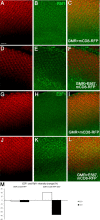
Fig 7. HPV16 E5, E6, E7 reduce the level of Drosophila Retinoblastoma protein Rbf1 resulting in an increase in E2F1.
Transgenes were expressed in the third instar larval eye discs using GMR-Gal4. mCD8RFP was used to mark the transgenes expression region and tissues were immunolabeled for Rbf1 and its interacting protein, E2F1. (A-C) Expression of mCD8RFP had no effect on Rbf1, which was equally present in mCD8RFP-expressing and non-expressing regions (B). (D-F) Co-expression of mCD8RFP +E5, E6, E7 led to reduction of Rbf1 (E). (G-I) Expression of mCD8RFP had no effect on E2F1 and was present in low levels in the nuclei (H). (J-L) Co-expression of mCD8RFP +E5, E6, E7 led to an increase in the level of E2F1 in nuclei of cells expressing the oncogenes (K). (M) Quantification of the level of E2F1 and Rbf1 showed a significant reduction of the Rbf1 (38%, n = 6 eye discs) and a significant increase of E2F1 (21%, n = 7 eye discs) in eye discs expressing mCD8RFP +E5, E6, E7 compared to eye discs expressing mCD8RFP. Scale bar represents 100 μm.
Fig 8
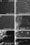
Fig 8. HPV16 E5, E6, and E7 oncoproteins induce the formation of long cellular protrusions.
Transgenes were expressed in the epithelium of the eye tissues using GMR-Gal4. mCD8RFP was used to mark the cell membranes in the GMR-Gal4 expression region. All eyes are from 42 hours after pupation. (A) Pupal eye expressing mCD8RFP exhibited an array of hexagonal ommatidia arranged in stereotypical pattern. (B) Co-expression of mCD8RFP+E5, E6, E7 had no effect on the ommatidia formation and organization. (C) Co-expression of mCD8RFP+E5, E6, E7+p35 did not perturb ommatidia development and arrangement. Co-expression of E5, E6, E7+hUBE3A+ mCD8RFP (D, D’) or co-expression of E5, E6, E7+hUBE3A+ mCD8RFP+p35 (E, E’) both resulted in loss of ommatidia differentiation. Pupal eye showed clusters of cells with rounded cell morphology. A subset of these cells were malignant and found at distant sites from the cluster (D, E). Some of these cells extended long cellular protrusions originating from opposing regions of the cell that linked the metastatic cells to each other or to the cells within the cluster (D’ and E’ magnified from boxes in D and E). Insets are digitally magnified 200%. Scale bar in A indicates 100 μm and in insets represents 50 μm.
Fig 9
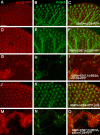
Fig 9. HPV16 oncogenes in cooperation with hUBE3A induce the transformation of epithelial cells to neuron-like cells.
Transgenes were expressed in the epithelium of the eye tissues using GMR-Gal4. mCD8RFP was used to mark the cell membranes in the GMR-Gal4 expression region. All eyes are from 42 hours after pupation and immunolabeled for axonal marker, Futsch. (A-C) Pupal eye expressing mCD8RFP exhibited a stereotype pattern of hexagonal array of ommatidia (A) from which only the photoreceptor neurons of each ommatidia project axons that fasciculate together and project as a single bundle towards the optic lobes of the brain (B). Co-expression of mCD8RFP+E5, E6, E7 (D-F) or co-expression of mCD8RFP+E5, E6, E7+p35 (J-L) had no effect on ommatidia differentiation and photoreceptor axonal projections. Pupal eyes co-expressing E5, E6, E7+hUBE3A+ mCD8RFP (G-I) or E5, E6, E7+hUBE3A+ mCD8RFP+p35 (M-O) showed no differentiated ommatidia but instead clusters of cells (G, M) with rounded cell morphology and cellular protrusions that expressed the axonal marker Futsch (H, N). The axon-like protrusions were developed by both the malignant cells and cells within the primary cluster (I, O). Scale bar represents 100 μm.

References

    1. Pesut E, Dukic A, Lulic L, Skelin J, Simic I, Milutin Gasperov N, et al.. Human Papillomaviruses-Associated Cancers: An Update of Current Knowledge. Viruses. 2021;13(11). doi: 10.3390/v13112234 - DOI - PMC - PubMed
    1. Combes JD, Franceschi S. Role of human papillomavirus in non-oropharyngeal head and neck cancers. Oral Oncol. 2014;50(5):370–9. doi: 10.1016/j.oraloncology.2013.11.004 - DOI - PubMed
    1. de Martel C, Plummer M, Vignat J, Franceschi S. Worldwide burden of cancer attributable to HPV by site, country and HPV type. Int J Cancer. 2017;141(4):664–70. doi: 10.1002/ijc.30716 - DOI - PMC - PubMed
    1. Munoz N, Bosch FX, de Sanjose S, Herrero R, Castellsague X, Shah KV, et al.. Epidemiologic classification of human papillomavirus types associated with cervical cancer. N Engl J Med. 2003;348(6):518–27. doi: 10.1056/NEJMoa021641 - DOI - PubMed
    1. Gillison ML, Koch WM, Capone RB, Spafford M, Westra WH, Wu L, et al.. Evidence for a causal association between human papillomavirus and a subset of head and neck cancers. J Natl Cancer Inst. 2000;92(9):709–20. doi: 10.1093/jnci/92.9.709 - DOI - PubMed

Publication types

MeSH terms

Substances