

Case Reports
doi: 10.4269/ajtmh.22-0441.
Print 2023 Jan 11.
Case Report: Poor Outcome Despite Aggressive Management in Pythium insidiosum Endophthalmitis
Affiliations
- PMID: 36509056
- PMCID: PMC9833092
- DOI: 10.4269/ajtmh.22-0441
Item in Clipboard
Case Reports
Case Report: Poor Outcome Despite Aggressive Management in Pythium insidiosum Endophthalmitis
Am J Trop Med Hyg.
.
Abstract
Pythium insidiosum is a parasitic oomycete that can cause keratitis and closely resembles fungus, both clinically and morphologically. It requires a trained microbiologist for its differentiation from fungal filaments and has poor response to antifungal therapy. We present a case of primary isolation of the organism from the vitreous cavity in a case of endophthalmitis. The infection spread quickly and involved all the ocular tissues. The eye had poor visual outcome despite a sequence of rapidly completed retinal and corneal surgeries along with initiation of anti-Pythium therapy.
Figures

(A) Slit-lamp picture of Pythium keratitis revealing the typical signs: ring infiltrate and guttering. (B) B-scan picture of day 1 depicting numerous vitreous echoes (C), which started resolving with appropriate anti-Pythium therapy.
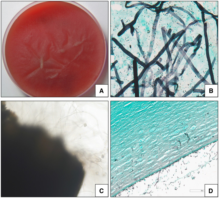
(A) Flat, adherent, velvety growth of P. insidiosum from a vitreous biopsy on blood agar. (B) Broad, aseptate, fungal-like filaments with characteristic ribbon-like folds of P. insidiosum on Gomori methenamine silver stain from growth on blood agar, seen under ×40 magnification by bright-field microscopy. (C) Zoospore formation of P. insidiosum at the margins of infected leaves in induction medium, seen under ×10 magnification by bright-field microscopy. (D) Histopathology showed cut-end and aseptate, ribbon-like filaments of Pythium in the posterior stroma and anterior chamber of the cornea on Grocott-Gomori methenamine silver stain under ×400 magnification.

(A) Slit-lamp picture of postoperative day 1 after therapeutic keratoplasty. Sutures are intact and the globe is formed. (B and C) B scan revealing decreases in vitreous echoes but the development of retinal detachment. (D) Slit-lamp picture upon the last follow-up. Most of the sutures are removed and the eye is going into phthisis.
References
-
- Sharma S Balne PK Motukupally SR Das S Garg P Sahu SK Arunasri K Manjulatha K Mishra DK Shivaji S , 2015. Pythium insidiosum keratitis. Cornea 34: 438–442. - PubMed
-
- Bagga B Kate A Mohamed A Sharma S Das S Mitra S , 2021. Successful strategic management of Pythium insidiosum keratitis with antibiotics. Ophthalmology 128: 169–172. - PubMed
Publication types
MeSH terms
LinkOut - more resources
Full Text Sources
