

Collagen is More Abundant and Structurally Altered in Lichen Sclerosus
- PMID: 36509210
- PMCID: PMC10038846
- DOI: 10.1016/j.urology.2022.11.036
Collagen is More Abundant and Structurally Altered in Lichen Sclerosus
Abstract
Objective: To test the hypothesis that genital skin and male urethra affected by lichen sclerosus (LS) has increased collagen content and altered collagen structure.
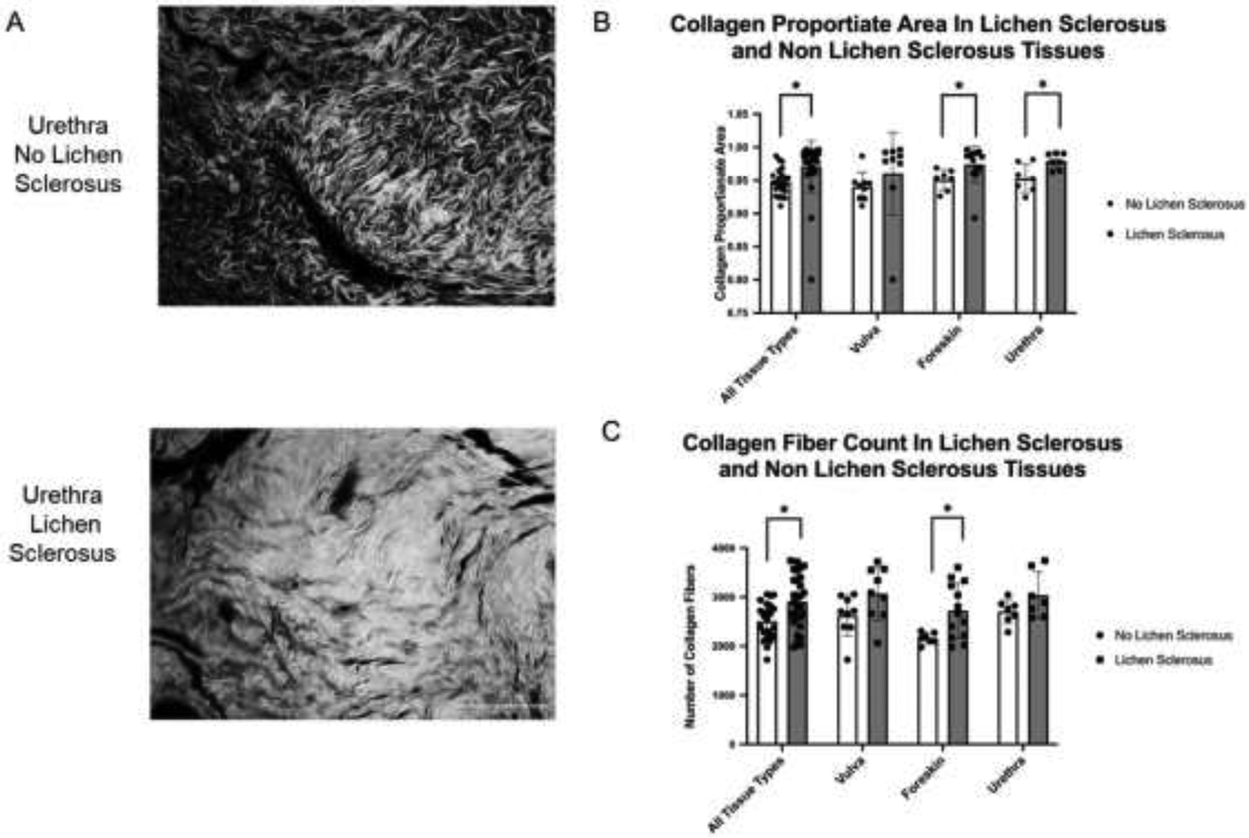
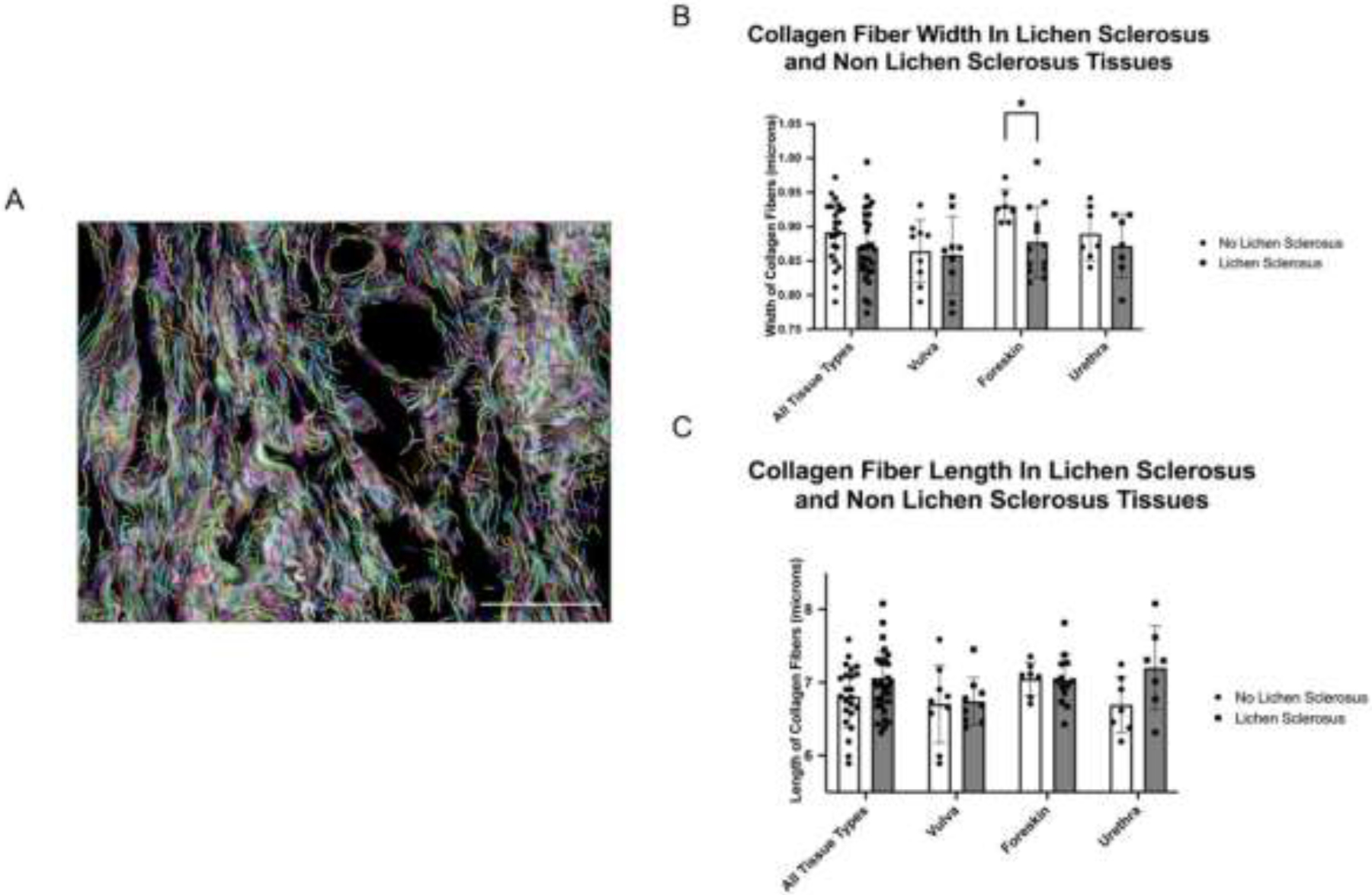
Methods: We used picrosirius red to stain and image collagen in human urethral, vulvar, and foreskin specimens with and without LS. Using Image J software, we quantified and compared (1) collagen content (using 2o metrics: collagen proportionate area [CPA] and collagen fiber count), (2) collagen fiber length and width, and (3) collagen structure using the texture analysis technique gray level co-localization matrix (GLCM) with respect to LS status and tissue type.
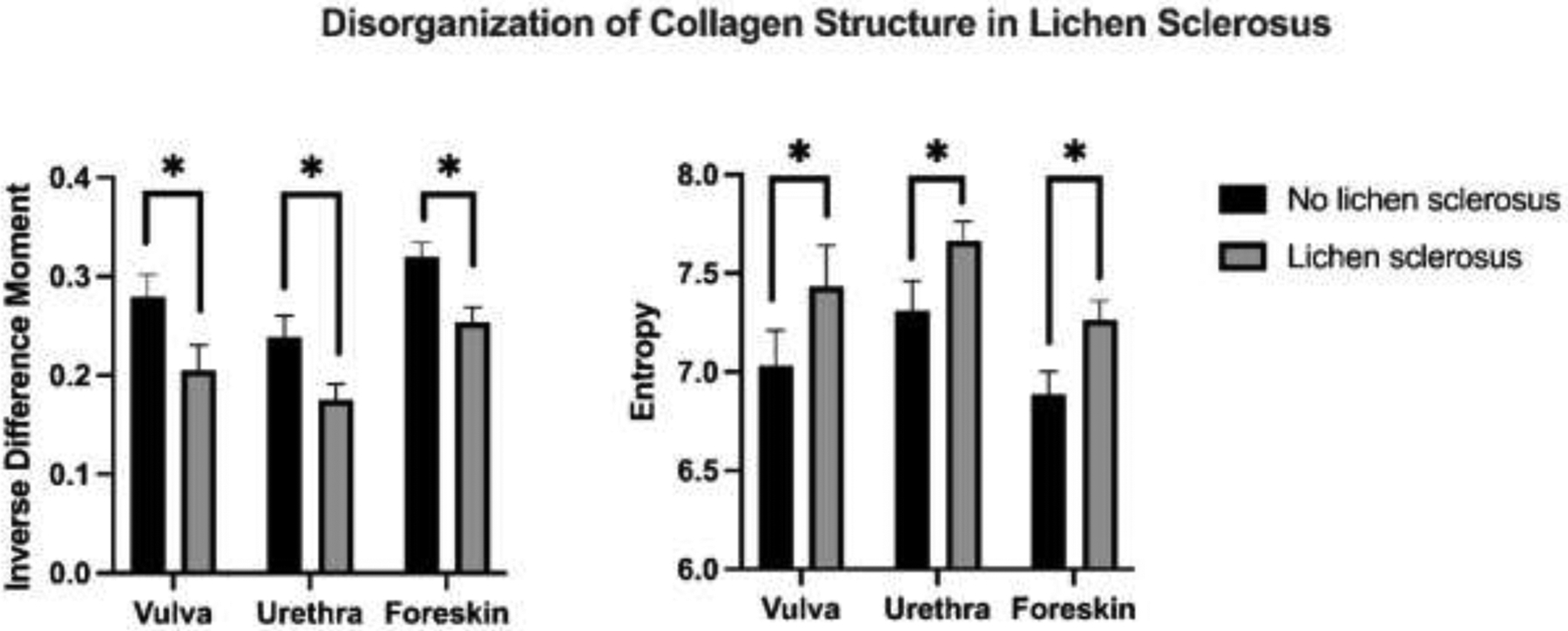
Results: We analyzed 23 LS specimens (vulva n=9, urethra n=7, foreskin n=7) and 29 non-LS specimens (vulva n=9, urethra n=7, foreskin n=13). Fiber count and CPA were significantly higher in all LS specimens compared to non-LS specimens (CPA: mean±SD 0.971±0.03 vs 0.948±0.02, P < .007; fiber count: mean±SD = 2906±127 vs 2509±78 fibers; P = .003). Collagen fiber width and length were similar with respect to LS status. GLCM analysis showed decreased inverse difference moment and increased entropy in LS tissues indicative of less homogeneous and more disorganized tissue structure (P<.001).
Conclusion: LS tissues have greater collagen content compared to non-LS tissues. Quantitative assessment of collagen organization, using GLCM, revealed less homogeneity and more disorganization of collagen in LS compared to non-LS tissues. Taken together, our findings suggest that alterations in physical tissue properties seen in LS may be due to both increased collagen abundance and altered structure.
Copyright © 2022 Elsevier Inc. All rights reserved.
Figures
Similar articles
-
Evaluation of genital self-image and sexual dysfunction in women with vulvar lichen planus or lichen sclerosus.J Psychosom Obstet Gynaecol. 2022 Jun;43(2):99-106. doi: 10.1080/0167482X.2020.1857359. Epub 2020 Dec 10. J Psychosom Obstet Gynaecol. 2022. PMID: 33297796
-
Unusual remodeling of the hyalinization band in vulval lichen sclerosus by type V collagen and ECM 1 protein.Clinics (Sao Paulo). 2015 May;70(5):356-62. doi: 10.6061/clinics/2015(05)09. Epub 2015 May 1. Clinics (Sao Paulo). 2015. PMID: 26039953 Free PMC article.
-
Langerhans cells in lichen sclerosus of the vulva and lichen sclerosus evolving in vulvar squamous cell carcinoma.Histol Histopathol. 2009 Mar;24(3):331-6. doi: 10.14670/HH-24.331. Histol Histopathol. 2009. PMID: 19130403
-
Vulvar lichen sclerosus and squamous cell carcinoma: a cohort, case control, and investigational study with historical perspective; implications for chronic inflammation and sclerosis in the development of neoplasia.Hum Pathol. 1998 Sep;29(9):932-48. doi: 10.1016/s0046-8177(98)90198-8. Hum Pathol. 1998. PMID: 9744309 Review.
-
Current treatment of lichen sclerosus and stricture.World J Urol. 2020 Dec;38(12):3061-3067. doi: 10.1007/s00345-019-03030-z. Epub 2019 Dec 5. World J Urol. 2020. PMID: 31807846 Review.
Cited by
-
Immune Dysregulation and Cellular Composition in Lichen Sclerosus Revealed by Integrative Epigenetic Analysis with Cell Type Deconvolution.J Inflamm Res. 2025 Jan 7;18:283-299. doi: 10.2147/JIR.S481324. eCollection 2025. J Inflamm Res. 2025. PMID: 39802516 Free PMC article.
-
Multi-omics analysis unveiled fibroblast-mediated pathogenesis in male genital lichen sclerosus.Cell Biosci. 2025 Jul 31;15(1):113. doi: 10.1186/s13578-025-01453-3. Cell Biosci. 2025. PMID: 40745572 Free PMC article.
References
-
- Granieri MA, Peterson AC, Madden-Fuentes RJ. Effect of Lichen Sclerosis on Success of Urethroplasty. Urol Clin North Am. 2017;44(1):77–86. - PubMed
-
- Fergus KB, Lee AW, Baradaran N, Cohen AJ, Stohr BA, Erickson BA, et al. Pathophysiology, Clinical Manifestations, and Treatment of Lichen Sclerosus: A Systematic Review. Urology. 2020;135:11–9. - PubMed
-
- Levy A, Browne B, Fredrick A, Stensland K, Bennett J, Sullivan T, et al. Insights into the Pathophysiology of Urethral Stricture Disease due to Lichen Sclerosus: Comparison of Pathological Markers in Lichen Sclerosus Induced Strictures vs Nonlichen Sclerosus Induced Strictures. J Urol. 2019;201(6):1158–63. - PubMed
-
- Querejeta R, Lopez B, Gonzalez A, Sanchez E, Larman M, Martinez Ubago JL, et al. Increased collagen type I synthesis in patients with heart failure of hypertensive origin: relation to myocardial fibrosis. Circulation. 2004;110(10):1263–8. - PubMed
Publication types
MeSH terms
Substances
Grants and funding
LinkOut - more resources
Full Text Sources
