. 2023 Jan:162:105462.
doi: 10.1016/j.neuint.2022.105462. Epub 2022 Dec 9.

Cerebral microvascular matrix metalloproteinase-3 (MMP3) contributes to vascular injury after stroke in female diabetic rats

Affiliations

Cerebral microvascular matrix metalloproteinase-3 (MMP3) contributes to vascular injury after stroke in female diabetic rats

Yasir Abdul et al. Neurochem Int. 2023 Jan.

Abstract

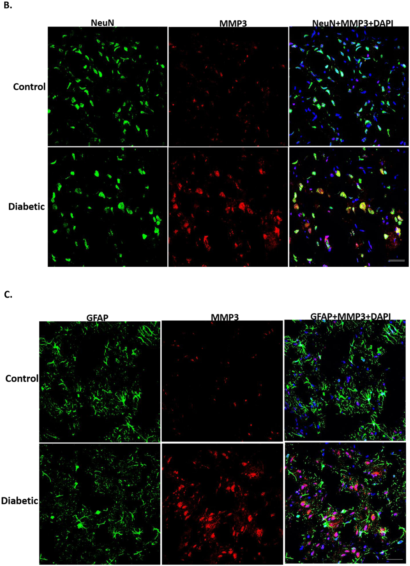
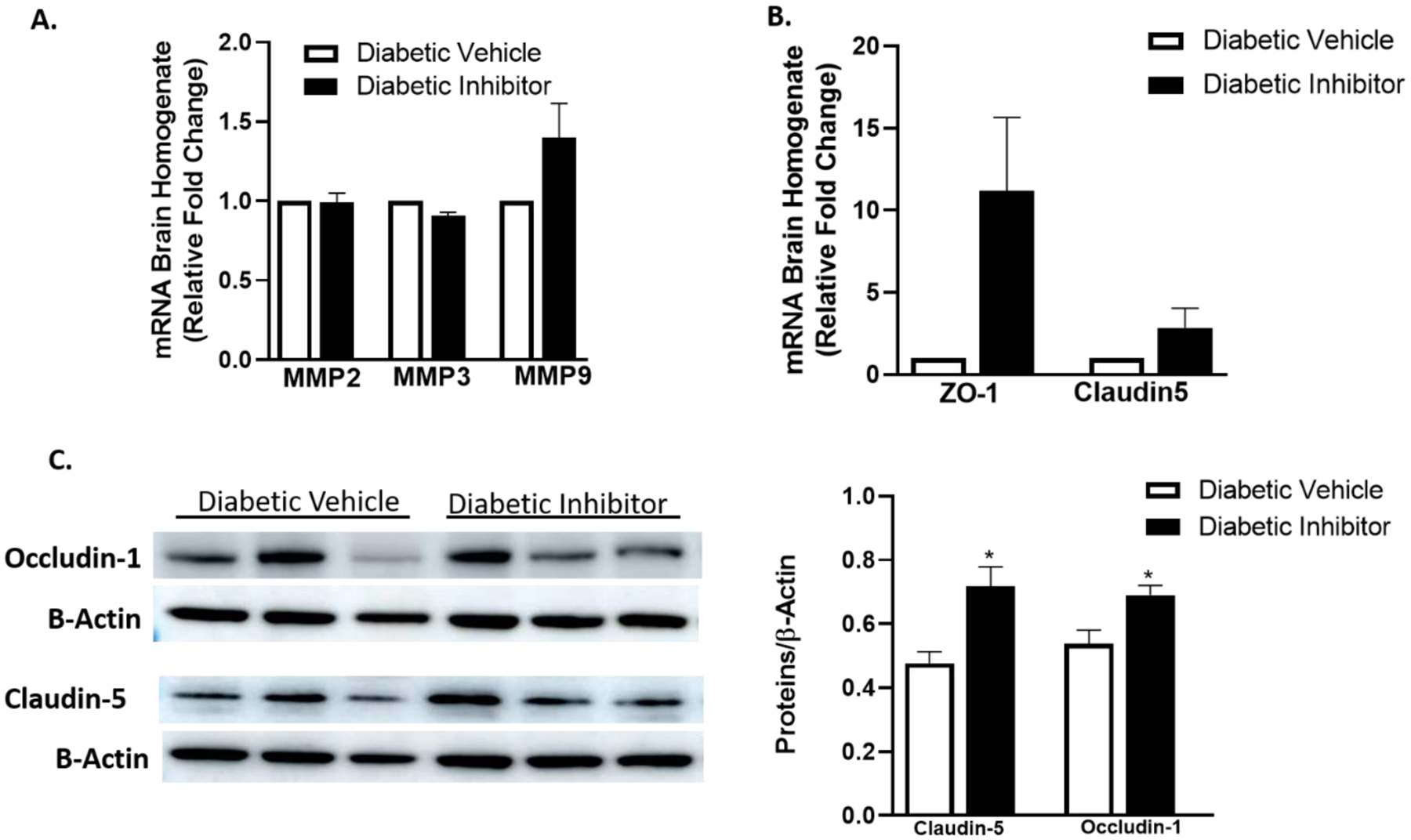
Diabetes exacerbates hemorrhagic transformation (HT) after stroke and worsens clinical outcomes. Female patients with diabetes are at a greater risk of stroke and worsened recovery. We have shown that activation of matrix metalloprotease 3 (MMP3) in hyperglycemic settings mediates HT in male rats. In light of our recent findings that diabetic female rats develop greater HT, the current study was designed to test the hypotheses that: 1) cerebral microvascular MMP3 activation contributes to poor functional outcomes and increased hemorrhagic transformations (HT) after ischemic stroke, and 2) MMP3 inhibition can improve functional outcomes in female diabetic rats. Female control and diabetic Wistar rats were subjected to 60 min of middle cerebral artery occlusion (MCAO). One cohort of diabetic animals received a single dose of MMP3 inhibitor (UK356618; 15 mg/kg; iv) or vehicle after reperfusion. Neurobehavioral outcomes, brain infarct size, edema, HT, and MMPs were measured in brain tissue. Diabetic rats had significant neurological deficits on Day 3 after stroke. MMP3 expression and enzyme activity were significantly increased in both micro and macro vessels of diabetic animals. MMP3 inhibition improved functional outcomes and reduced brain edema and HT scores. In conclusion, cerebral endothelial MMP3 activation to vascular injury in female diabetic rats. Our findings identify MMP3 as a potential therapeutic target in diabetic stroke.

Keywords: Brain; Diabetes; Endothelial cells; MMP3; Stroke.


Conflict of interest statement

Declaration of competing interest The authors declare that they have no known competing financial interests or personal relationships that could have appeared to influence the work reported in this paper.

Figures

Fig. 1
Fig. 1
Schematic timeline and description of the experimental design, animal numbers per group, and mortality rate.
Fig. 2
Fig. 2
Ischemic stroke in female diabetic animals worsens behavioral outcomes and increases MMP3 in brain microvessels. Measurement of the composite score (A) and adhesive removal test (B) was performed 3 days after ischemic stroke in control and diabetic female rats. (*, p<0.05, vs controls group; n=12). Expression of MMP3 protein (C) and MMP3 enzyme activity (D) in isolated brain microvessels was significantly higher in diabetic animals compared to control (*, p<0.05; n=5–6 in each group).
Fig. 3
Fig. 3
Ischemic stroke increased the expression of MMP genes in the brain. (A) mRNA expression of MMP2, MMP3, and MMP9 measured in the ipsilateral side of the brain was robustly increased in diabetic animals compared to control animals. (B) MMP3 and MMP9 were also expressed in brain macrovessels (MCA) of diabetic animals compared to control animals (n= 4–6 in each group).
Fig. 4
Fig. 4
Representative images of immunofluorescence showing MMP3 expression in the cortex of the ischemic hemisphere 3 days after ischemic stroke. MMP3 immunoreactivity was greater in diabetic animals. MMP3 is colocalized with endothelial cell marker CD31 (A) and neuronal marker NeuN (B). While MMP3 expression was more around astrocyte cell marker GFAP (C). Images were captured at 20x magnification and the scale bar is 25 μm (n=4 in each group).
Fig. 4
Fig. 4
Representative images of immunofluorescence showing MMP3 expression in the cortex of the ischemic hemisphere 3 days after ischemic stroke. MMP3 immunoreactivity was greater in diabetic animals. MMP3 is colocalized with endothelial cell marker CD31 (A) and neuronal marker NeuN (B). While MMP3 expression was more around astrocyte cell marker GFAP (C). Images were captured at 20x magnification and the scale bar is 25 μm (n=4 in each group).
Fig. 5
Fig. 5
Hypoxia activates MMPs in female HBMECs in diabetic conditions. Hypoxia (CoCl2) increased the mRNA expression of MMP2, MMP3, and MMP9 (A) in both normal (NG) and diabetic (HG+ P; B) conditions. interestingly, this increase was much higher in diabetic conditions compared to normal conditions (n=4–5). MMP3 enzyme activity (C) measured by FRET assay and MMP2 and MMP9 (D & E) activity measured by zymogram was significantly increased in media collected from cells in diabetic conditions, however, hypoxia did not increase it further (n=4).
Fig. 6
Fig. 6
Inhibition of MMP3 reduces the neurovascular injury in female diabetic animals subjected to ischemic stroke. Female diabetic rats were subjected to 60 min of MCAO and treated with vehicle or MMP3 inhibitor UK356618 (15 mg/kg; i.v.) at reperfusion. After 72 h of reperfusion, animals were sacrificed to measure the neurovascular injury. (A) Representative images showing edema and infarct (arrows indicate the hemorrhages). (B) Treatment with an MMP3 inhibitor did not show any difference in infarct size. (C) Edema was significantly reduced in animals treated with MMP3 inhibitor (***, p<0.001). (D) Hemorrhagic transformation (HT) index was significantly reduced in animals treated with MMP3 inhibitor compared to vehicle-treated animals (*, p<0.05; n=7 in each group).
Fig. 7
Fig. 7
Inhibition of MMP3 improves functional outcomes in female diabetic animals after ischemic stroke. (A) The composite score measured on Day 3 post-stroke was improved in animals treated with an MMP3 inhibitor. (B) Adhesive removal time (ART) was significantly reduced in animals treated with MMP3 inhibitor (*p < 0.05; n=7–9 in each group).
Fig. 8
Fig. 8
Inhibition of MMP3 after ischemic stroke improves the expression of tight junction proteins in female diabetic animals. (A) mRNA expression of MMP2, MMP3, and MMP9 was not different in the ischemic side of the brain between the vehicle and MMP3 inhibitor-treated animals. (B) mRNA expression of ZO-1 and claudin5 in the ischemic side of the brain was higher in animals treated with an MMP3 inhibitor. (C) Tight junction proteins occludin1 and claudin5 were also significantly higher in the ischemic side of the brain in animals treated with MMP3 inhibitor (*, p<0.05, n=6–9 in each group).

References

    1. Abdul Y, Abdelsaid M, Li W, Webb RC, Sullivan JC, Dong G, and Ergul A 2019. Inhibition of Toll-Like Receptor-4 (TLR-4) Improves Neurobehavioral Outcomes After Acute Ischemic Stroke in Diabetic Rats: Possible Role of Vascular Endothelial TLR-4. Mol Neurobiol 56(3): 1607–1617. doi: 10.1007/s12035-018-1184-8. - DOI - PMC - PubMed
    1. Abdul Y, Li W, Vargas JD, Grant E, He L, Jamil S, and Ergul A 2020. Diabetes-related sex differences in the brain endothelin system following ischemia in vivo and in human brain endothelial cells in vitro. Can J Physiol Pharmacol 98(9): 587–595. doi: 10.1139/cjpp-2019-0630. - DOI - PMC - PubMed
    1. Appelros P, Stegmayr B, and Terent A 2009. Sex differences in stroke epidemiology: a systematic review. Stroke 40(4): 1082–1090. doi: 10.1161/STROKEAHA.108.540781. - DOI - PubMed
    1. Baird TA, Parsons MW, Phan T, Butcher KS, Desmond PM, Tress BM, Colman PG, Chambers BR, and Davis SM 2003. Persistent poststroke hyperglycemia is independently associated with infarct expansion and worse clinical outcome. Stroke 34(9): 2208–2214. doi: 10.1161/01.STR.0000085087.41330.FF. - DOI - PubMed
    1. Bejot Y, and Giroud M 2010. Stroke in diabetic patients. Diabetes Metab 36 Suppl 3: S84–87. doi: 10.1016/S1262-3636(10)70472-9. - DOI - PubMed

Publication types

Substances