. 2023 Jun;44(6):1161-1174.
doi: 10.1038/s41401-022-01031-0. Epub 2022 Dec 12.

SGLT2 inhibitor empagliflozin promotes revascularization in diabetic mouse hindlimb ischemia by inhibiting ferroptosis

Affiliations

SGLT2 inhibitor empagliflozin promotes revascularization in diabetic mouse hindlimb ischemia by inhibiting ferroptosis

Jing-Xuan Han et al. Acta Pharmacol Sin. 2023 Jun.

Abstract

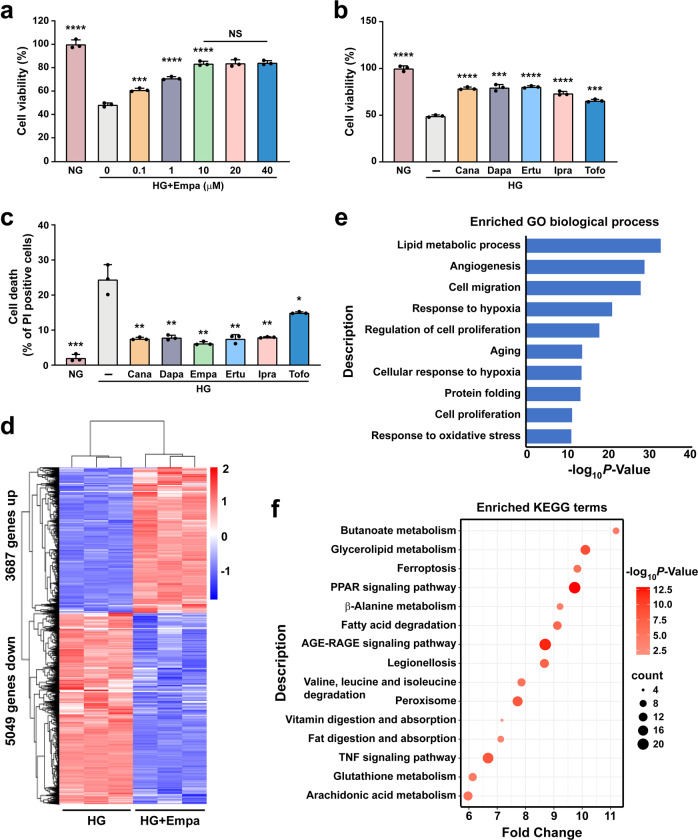
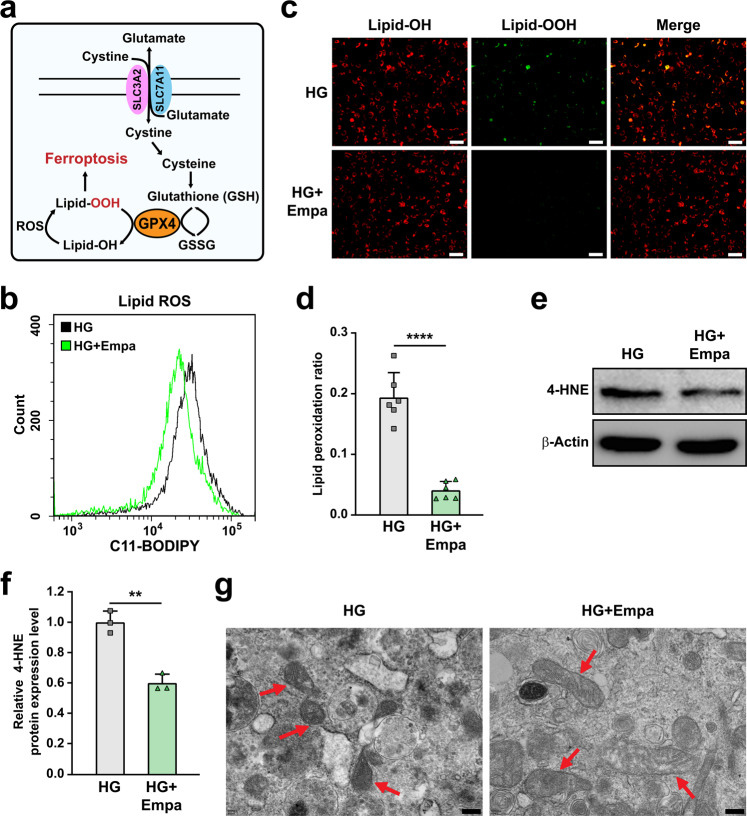
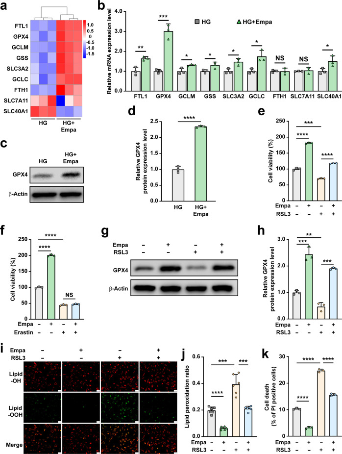
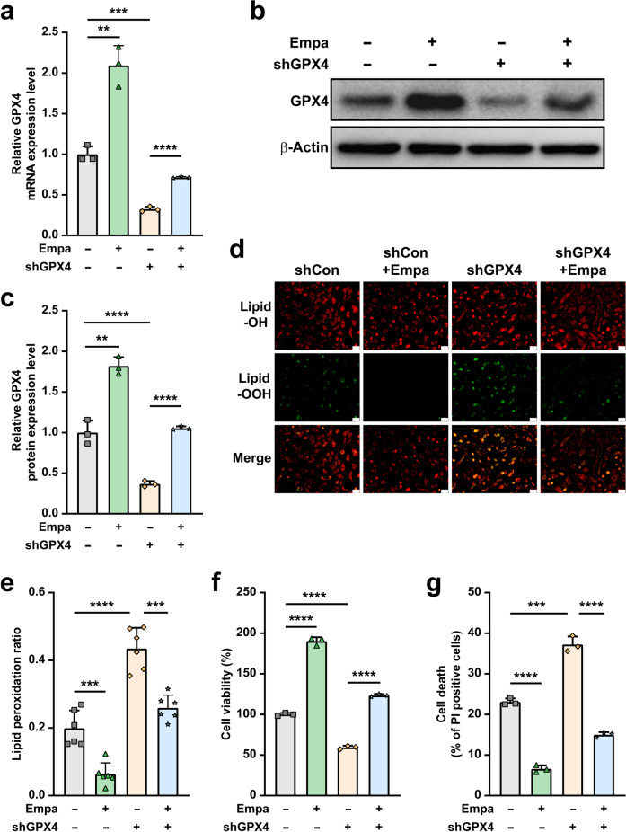
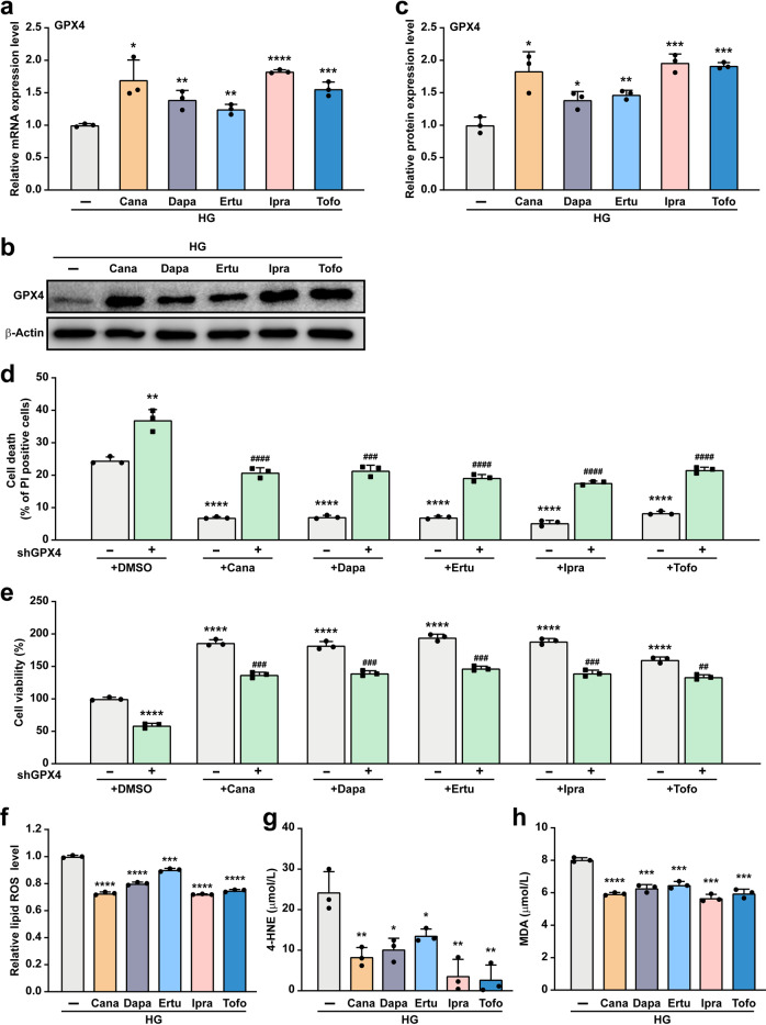
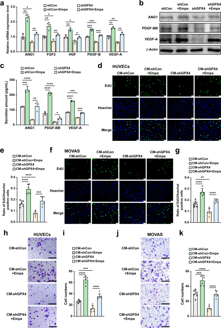
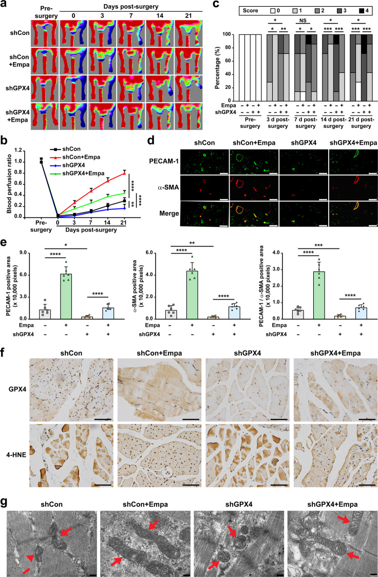
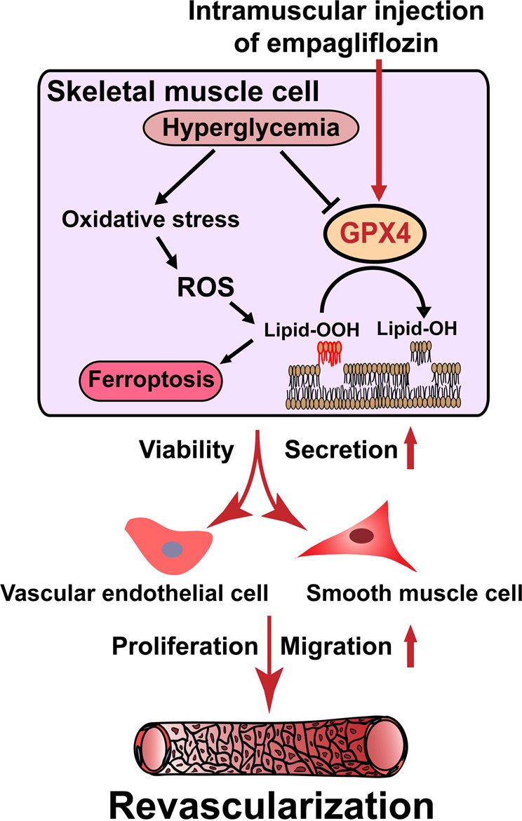
Gliflozins are known as SGLT2 inhibitors, which are used to treat diabetic patients by inhibiting glucose reabsorption in kidney proximal tubules. Recent studies show that gliflozins may exert other effects independent of SGLT2 pathways. In this study we investigated their effects on skeletal muscle cell viability and paracrine function, which were crucial for promoting revascularization in diabetic hindlimb ischemia (HLI). We showed that treatment with empagliflozin (0.1-40 μM) dose-dependently increased high glucose (25 mM)-impaired viability of skeletal muscle C2C12 cells. Canagliflozin, dapagliflozin, ertugliflozin, ipragliflozin and tofogliflozin exerted similar protective effects on skeletal muscle cells cultured under the hyperglycemic condition. Transcriptomic analysis revealed an enrichment of pathways related to ferroptosis in empagliflozin-treated C2C12 cells. We further demonstrated that empagliflozin and other gliflozins (10 μM) restored GPX4 expression in high glucose-treated C2C12 cells, thereby suppressing ferroptosis and promoting cell viability. Empagliflozin (10 μM) also markedly enhanced the proliferation and migration of blood vessel-forming cells by promoting paracrine function of skeletal muscle C2C12 cells. In diabetic HLI mice, injection of empagliflozin into the gastrocnemius muscle of the left hindlimb (10 mg/kg, every 3 days for 21 days) significantly enhanced revascularization and blood perfusion recovery. Collectively, these results reveal a novel effect of empagliflozin, a clinical hypoglycemic gliflozin drug, in inhibiting ferroptosis and enhancing skeletal muscle cell survival and paracrine function under hyperglycemic condition via restoring the expression of GPX4. This study highlights the potential of intramuscular injection of empagliflozin for treating diabetic HLI.

Keywords: diabetic hindlimb ischemia; empagliflozin; ferroptosis; revascularization; therapeutic angiogenesis.


Conflict of interest statement

All authors declare no competing interests. The results of this study have been patented in China (No. ZL202010701077.7).

Figures

Fig. 1
Fig. 1. Gliflozins promote skeletal muscle cells survival under hyperglycemia.
a Cell viability of C2C12 cells treated with indicated concentration of empagliflozin. b Cell viability of C2C12 cells treated with 10 μM gliflozins. c Cell death rate of C2C12 cells treated with 10 μM gliflozins, as examined using PI staining and flow cytometry. d RNA-sequencing analysis of C2C12 treated with 10 μM empagliflozin. Heat-map represents the result of clustering analysis of differentially expressed genes with fold-change ≥1.5 and P-value < 0.05. e GO analysis of differentially expressed genes with fold-change ≥1.5 and P-value < 0.05. Enriched GO biological processes were identified and listed according to their enrichment scores (-log10 P-value). f KEGG enrichment analysis of differentially expressed genes with fold-change ≥1.5 and P-value < 0.05. C2C12 cells cultured under hyperglycemia were used as control. All experiments were performed under hyperglycemia unless further indicated. Data were presented as mean ± SD (n = 3). NG normoglycemia, HG hyperglycemia, NS not significant, *P < 0.05; **P < 0.01; ***P < 0.001; ****P < 0.0001.
Fig. 2
Fig. 2. Empagliflozin inhibits ferroptosis in skeletal muscle cells under hyperglycemia.
a Schematic diagram of molecular mechanism of ferroptosis. b Lipid ROS level in empagliflozin-treated C2C12 cells, as examined using C11-BODIPY staining and flow cytometry. c, d Lipid peroxidation ratio in empagliflozin-treated C2C12 cells, as analyzed using C11-BODIPY staining. Representative images (c; scale bars: 100 μm) and quantification results (d; n = 6) were shown. e, f 4-HNE level in C2C12 cells treated with empagliflozin, as examined using Western blotting. Representative images (e) and quantification results (f) were shown. β-Actin was used as loading control. g Transmission electron microscopy images of the mitochondria in C2C12 cells treated with empagliflozin. Red arrows: mitochondria; scale bars: 200 nm. All experiments were performed under hyperglycemia. Data were presented as mean ± SD (n = 3, unless further indicated). HG hyperglycemia, Empa: 10 μM empagliflozin; **P < 0.01; ****P < 0.0001.
Fig. 3
Fig. 3. Empagliflozin inhibits hyperglycemia-induced ferroptosis by enhancing GPX4 expression.
a Heatmap showing ferroptosis-related genes which are differently expressed genes in C2C12 cells cultured under hyperglycemia and treated with 10 μM empagliflozin obtained by RNA-seq. Values are scaled as indicated (1.0 to −1.5). b mRNA expression levels of ferroptosis-related genes in C2C12 cells cultured under hyperglycemia and treated with 10 μM empagliflozin. c, d GPX4 protein expression in C2C12 cells cultured under hyperglycemia and treated with 10 μM empagliflozin, as examined using Western blotting. Representative images (c) and quantification results (d) were shown. Cell viability of C2C12 cells treated with 10 μM empagliflozin and 100 nM RSL3 (e) or 500 nM erastin (f). g, h GPX4 protein expression in C2C12 cells treated with 10 μM empagliflozin and 100 nM RSL3, as examined using Western blotting. Representative images (g) and quantification results (h) were shown. i, j Lipid peroxidation ratio in C2C12 cells treated with 10 μM empagliflozin and 100 nM RSL3, as analyzed using C11-BODIPY staining. Representative images (i; scale bars: 100 μm) and quantification results (j; n = 6) were shown. k Cell death rate of C2C12 cells treated with 10 μM empagliflozin and 100 nM RSL3 as examined using PI staining and flow cytometry. β-Actin was used for qRT-PCR normalization and as Western blotting loading control. All experiments were performed under hyperglycemia. Data were presented as mean ± SD (n = 3, unless further indicated). HG hyperglycemia, NS not significant; *P < 0.05; **P < 0.01; ***P < 0.001; ****P < 0.0001.
Fig. 4
Fig. 4. Empagliflozin inhibits ferroptosis under hyperglycemia through GPX4.
GPX4 mRNA (a) and protein (b, c) expression in GPX4-knocked down C2C12 cells treated with 10 μM empagliflozin, as examined using qRT-PCR and Western blotting, respectively. Representative images (b) and quantification results (c) were shown. β‐Actin was used for qRT-PCR normalization and as Western blotting loading control. d, e Lipid peroxidation ratio in GPX4-knocked down C2C12 cells treated with 10 μM empagliflozin, as analyzed using C11-BODIPY staining. Representative images (d; scale bars: 100 μm) and quantification results (e; n = 6) were shown. f Cell viability in GPX4-knocked down C2C12 cells treated with 10 μM empagliflozin. g Cell death rate in GPX4-knocked down C2C12 cells treated with 10 μM empagliflozin, as examined using PI staining and flow cytometry. All experiments were performed under hyperglycemia. Data were presented as mean ± SD (n = 3, unless further indicated). **P < 0.01; ***P < 0.001; ****P < 0.0001.
Fig. 5
Fig. 5. Gliflozins inhibit ferroptosis under hyperglycemia through GPX4.
mRNA (a) and protein (b, c) expression of GPX4 in C2C12 cells treated with 10 μM of indicated gliflozins, as examined using qRT-PCR and Western blotting, respectively. Representative images (b) and quantification results (c) were shown. β‐Actin was used for qRT-PCR normalization and as Western blotting loading control. d Cell death rate in GPX4-knocked down C2C12 cells treated with 10 μM of indicated gliflozins, as examined using PI staining and flow cytometry. e Cell viability in GPX4-knocked down C2C12 cells treated with 10 μM of indicated gliflozins. f Lipid ROS level in C2C12 cells treated with 10 μM of indicated gliflozins, as examined using C11-BODIPY staining and flow cytometry. 4-HNE (g) and MDA (h) levels in C2C12 cells treated with 10 μM of indicated gliflozins, as analyzed using ELISA and Lipid Peroxidation MDA Assay Kit, respectively. All experiments were performed under hyperglycemia. Data were presented as mean ± SD (n = 3). HG hyperglycemia; *P < 0.05; **P < 0.01; ***P < 0.001; ****P < 0.0001 versus shCon; while #P < 0.05; ##P < 0.01; ###P < 0.001; ####P < 0.0001 versus shGPX4.
Fig. 6
Fig. 6. Empagliflozin promotes blood vessel-forming cells proliferation and migration by enhancing skeletal muscle cells paracrine function in a GPX4-dependent manner.
mRNA (a) and protein (b) expression of angiogenic factors in GPX4-knocked down C2C12 cells treated with 10 μM empagliflozin, as examined using qRT-PCR and Western blotting, respectively. β‐Actin was used for qRT-PCR normalization and as Western blotting loading control. c Secreted amount of ANG1, PDGF-BB and VEGF-A in the culture medium of C2C12 cells treated with 10 μM empagliflozin, as analyzed using ELISA. Proliferation potential of HUVECs (d, e) and MOVAS cells (f, g) cultured with indicated conditioned media collected from GPX4-knocked down C2C12 cells treated with 10 μM empagliflozin, as examined using EdU incorporation assay. Representative images (d and f; scale bars: 100 μm) and quantification results (e and g; n = 6) were shown. Migration potential of HUVECs (h, i) and MOVAS cells ( j, k) cultured with indicated conditioned media, as analyzed using transwell migration assay. Representative images (h and j; scale bars: 200 μm) and quantification results (i and k; n = 6) were shown. All experiments were performed under hyperglycemia. Data were presented as mean ± SD (n = 3, unless further indicated). CM-shCon and CM-shGPX4: conditioned media obtained from shCon-transfected or GPX4-knocked down C2C12 cells; CM-shCon+Empa and CM-shGPX4+Empa: conditioned media obtained from shCon- or shGPX4-transfected C2C12 cells treated with 10 μM empagliflozin. *P < 0.05; **P < 0.01; ***P < 0.001; ****P < 0.0001.
Fig. 7
Fig. 7. Empagliflozin enhances revascularization in diabetic HLI mice by inhibiting ferroptosis through GPX4.
a, b Blood perfusion in the ischemic hindlimbs of diabetic HLI mice intramuscularly injected with empagliflozin (10 mg/kg body weight) and shCon or shGPX4 vectors at indicated time points. Representative images (a) and quantification data of blood perfusion ratio were shown (b; n = 7). c Morphological assessment of ischemic hindlimbs in diabetic HLI mice intramuscularly injected with empagliflozin (10 mg/kg body weight) and shCon or shGPX4 vectors at indicated time points (n = 7). d, e Immunofluorescence against PECAM-1 (green) and α-SMA (red) in ischemic hindlimbs tissue of diabetic HLI mice intramuscularly injected with empagliflozin (10 mg/kg body weight) and shCon or shGPX4 vectors at day 21 after surgery. Representative images (d; scale bars: 50 μm) and quantification results (e) were shown. f Representative images of immunohistochemical staining against GPX4 and 4-HNE in ischemic hindlimbs tissues of diabetic HLI mice intramuscularly injected with empagliflozin (10 mg/kg body weight) and shCon or shGPX4 vectors at day 21 after surgery (scale bars: 50 μm). g Transmission electron microscopy images of the mitochondria in the ischemic hindlimbs tissues of diabetic HLI mice intramuscularly administered with empagliflozin (10 mg/kg body weight) and shCon or shGPX4 vectors at day 21 after surgery. Red arrows: mitochondria; scale bars: 200 nm. Data were presented as mean ± SD (n = 6). NS not significant; *P < 0.05; **P < 0.01; ***P < 0.001; ****P < 0.0001.
Fig. 8
Fig. 8. Schematic diagram showing the mechanism of intramuscularly-injected empagliflozin in enhancing therapeutic angiogenesis.
Empagliflozin inhibits hyperglycemia-induced ferroptosis in skeletal muscle cells by enhancing GPX4 expression, thus promoting revascularization in diabetic HLI mice.

References

    1. Washburn WN. Development of the renal glucose reabsorption inhibitors: a new mechanism for the pharmacotherapy of diabetes mellitus type 2. J Med Chem. 2009;52:1785–94. doi: 10.1021/jm8013019. - DOI - PubMed
    1. Neumiller JJ, White JR, Jr., Campbell RK. Sodium-glucose co-transport inhibitors: progress and therapeutic potential in type 2 diabetes mellitus. Drugs. 2010;70:377–85. doi: 10.2165/11318680-000000000-00000. - DOI - PubMed
    1. Isaji M. Sglt2 inhibitors: molecular design and potential differences in effect. Kidney Int Suppl. 2011;120:S14-9. - PubMed
    1. Neal B, Perkovic V, Mahaffey KW, de Zeeuw D, Fulcher G, Erondu N, et al. Canagliflozin and cardiovascular and renal events in type 2 diabetes. N Engl J Med. 2017;377:644–57.. doi: 10.1056/NEJMoa1611925. - DOI - PubMed
    1. Wiviott SD, Raz I, Bonaca MP, Mosenzon O, Kato ET, Cahn A, et al. Dapagliflozin and cardiovascular outcomes in type 2 diabetes. N Engl J Med. 2019;380:347–57.. doi: 10.1056/NEJMoa1812389. - DOI - PubMed

MeSH terms