. 2023 Apr;19(4):460-467.
doi: 10.1038/s41589-022-01206-0. Epub 2022 Dec 12.

Designer installation of a substrate recruitment domain to tailor enzyme specificity

Affiliations

Designer installation of a substrate recruitment domain to tailor enzyme specificity

Rodney Park et al. Nat Chem Biol. 2023 Apr.

Abstract

Promiscuous enzymes that modify peptides and proteins are powerful tools for labeling biomolecules; however, directing these modifications to desired substrates can be challenging. Here, we use computational interface design to install a substrate recognition domain adjacent to the active site of a promiscuous enzyme, catechol O-methyltransferase. This design approach effectively decouples substrate recognition from the site of catalysis and promotes modification of peptides recognized by the recruitment domain. We determined the crystal structure of this novel multidomain enzyme, SH3-588, which shows that it closely matches our design. SH3-588 methylates directed peptides with catalytic efficiencies exceeding the wild-type enzyme by over 1,000-fold, whereas peptides lacking the directing recognition sequence do not display enhanced efficiencies. In competition experiments, the designer enzyme preferentially modifies directed substrates over undirected substrates, suggesting that we can use designed recruitment domains to direct post-translational modifications to specific sequence motifs on target proteins in complex multisubstrate environments.


Conflict of interest statement

Competing Interests Statement

The authors declare no competing financial interests.

Figures

Extended Data Fig. 1.
Expanded set of timepoints for substrate competition assay for target peptide (red) and off-target (light blue) at 1 (A), 10 (B), and 100 (C) μM peptide substrate (2, 20, and 200 μM total peptide respectively). Raw data points are represented by black dots superimposed on plots. Data bars present mean values +/− SEM across three distinct reactions (n=3, frequently hidden under data points). Error bars are centered on the mean. (D) Exact timepoints collected for 1, 10, and 100 μM. Timepoint 2 is displayed in Fig. 4 in main text and t1 at 100 μM is used in competition reaction mathematical modeling.
Extended Data Fig. 2
Full computational model diagram. (A) Single, directed substrate simulation, where substrate can bind to the active site and peptide binding site. (B) Multi-substrate (directed and undirected competition) reaction simulation diagram.
Extended Data Fig. 3.
Substrate diversity assay. (A) Diagram of assay procedure. Briefly, kit components were combined with genes encoding peptides. After IVT incubation, the crude mixture was split, respective enzyme added, and reactions were run at 30°C for 15 minutes. Samples were prepped and run for MALDI-TOF-MS analysis. (B) Table of IVT peptide substrates tested and their corresponding conversions. SH3-588 largely reached completion for most peptides tested (marked with 100%); remaining substrate peaks were hardly above noise (IVT peptides 1-9). IVT peptides 1-10 were analyzed by MALDI-TOF-MS. Error values indicate +/− SEM across three distinct replicates (n=3) for IVT peptides 1-8 and two distinct replicates (n=2) for IVT peptides 9 and 10 centered on the mean.
Fig. 1.
Fig. 1.
General scaffolding diagrams and Rosetta design approach. (A) Flexible scaffolding approach used by our naïve fusion enzyme and Src family kinases. (B) Diagram of a specific interaction between a recruitment domain and promiscuous enzyme, similar to how many RiPPs enzymes are constructed. RS indicates the recognition sequence on the peptide that is bound by the peptide binding domain. (C) Overview of the Rosetta design pipeline. (D) Multistage Rosetta Scripts protocol (Design protocol in SI “dock_design_sh3.xml”) and follow-up design steps.
Fig. 2.
Fig. 2.
Structural characterization and background activity of designed enzymes. (A) Conversion of DOPA to methylated DOPA by COMT. (B) Plot of rate vs. [substrate] for WT COMT, naïve fusion, SH3-588, and SH3-003 for an undirected peptide substrate (1). All 3 enzymes have similar catalytic parameters with undirected substrate indicating that catalytic activity has not been compromised by the design process. The substrate residue, L-DOPA (YOH), is underlined. Rates are presented as points calculated from slopes of best fit calculated across 3 distinct reactions (n=3). Error bars are +/− SE of best fit centered on the calculated rate (example shown in Supplementary Fig. 3 A–D). (C) Crystal structure of SH3-588 (blue, PDB code: 7UD6) structurally aligned to its design model (gray). SAH (green) was observed in the crystal structure. The APP12 peptide is modeled bound to the SH3 domain (pink) to show relative placement near the active site. (D) Alignment of interface residues between SH3 and COMT domains showing packing at the interface is very similar in the crystal structure and design model. (E) Substrate peptide modeling on the crystal structure (a short peptide 7 (teal); medium peptide 2 (pink); and misdirected peptide 3 (orange)) indicating stretch of residues from peptide binding domain to active site. A magnesium ion (lime green) indicates the location of the active site but is not present in the SH3-588 crystal structure or during modeling.
Fig. 3.
Fig. 3.
Steady state kinetic analysis of the designed enzymes. The recognition sequence (RS) and L-DOPA substrate residue (YOH) are underlined in peptides. (A) Plot of rate vs. [substrate] for WT COMT, naïve fusion, SH3-588, and SH3-003 for a directed substrate (2). Boxed area indicates points that were not fit for the naïve fusion and suggest direct binding of substrate to the active site. (B) Plot of rate vs. [substrate] for WT COMT, naïve fusion, SH3-588, and SH3-003 for N-terminal misdirected substrate (3). (C) Plot of rate vs. [substrate] for SH3-588 for variable affinity recognition sequences (4,5,1). (D) Plot of rate vs. [substrate] for SH3-588 for variable length substrates (6,2,7). Rates are presented as points calculated from slopes of best fit calculated across 3 distinct reactions (n=3). Error bars are +/− SE of best fit centered on the calculated rate (example shown in Supplementary Fig. 3 A–D).
Fig. 4.
Fig. 4.
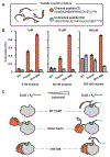
Substrate competition experiment. (A) Diagram showing competing substrates (peptides 2 and S2). (B) Percent conversion of directed and undirected peptides in competition reactions at 1, 10, and 100 μM (2, 20, and 200 μM total peptide) by indicated enzyme. Raw data points are represented by black dots superimposed on plots. Data bars present mean values +/− SEM across three distinct reactions (n=3). Error bars are centered on the mean. (C) Model of alternative modes of interaction between the enzymes and substrate at varying concentrations of substrate. KMActive site is the affinity (KM) of substrate binding directly to the active site.
Fig. 5.
Fig. 5.
Numerical modeling of competing substrates reacting with the engineered enzymes. (A) In the numerical model, directed substrates (i.e. containing the poly-proline recognition sequence) engage the SH3 domain and then exist in an equilibrium between an open and closed conformation (equilibrium constant = Kopen). In the closed form, the DOPA residue is bound to the active site and is available for catalysis. The fraction of bound peptide in the open conformation is referred to as fraction_open, which is directly related to Kopen. Undirected peptide can only engage the active site when the directed peptide is the open state, or the enzyme is not bound to directed peptide. Varying fraction open in the numerical model changes the accessibility of the active site to undirected peptides and changes the predicted specificity during a simulated reaction with directed and undirected peptide (the full mathematical model is presented in Extended Data Fig. 2). (B) A comparison of simulated results (hashed bars) and experimental results (solid bars) for a competitive reaction in which directed and undirected peptide are both at a concentration of 100 μM. Numerical simulations performed with different values assigned to fraction_open indicate that directed peptide is in the open conformation ~10% of the time when bound to SH3-588 and ~30% of the time when bound to the naïve fusion. Raw data points are represented by black dots superimposed on plots. Data bars present mean values +/− SEM across three distinct reactions (n=3). Error bars are centered on the mean. Hashed bars are a single computational result.

Similar articles

Cited by

References

    1. Ho SH & Tirrell DA Enzymatic Labeling of Bacterial Proteins for Super-resolution Imaging in Live Cells. ACS Cent Sci 5, 1911–1919 (2019). - PMC - PubMed
    1. Chen I, Howarth M, Lin W & Ting AY Site-specific labeling of cell surface proteins with biophysical probes using biotin ligase. Nat Methods 2, 99–104 (2005). - PubMed
    1. Bhattacharyya RP, Reményi A, Yeh BJ & Lim WA Domains, motifs, and scaffolds: The role of modular interactions in the evolution and wiring of cell signaling circuits. Annual Review of Biochemistry vol. 75 655–680 Preprint at 10.1146/annurev.biochem.75.103004.142710 (2006). - DOI - PubMed
    1. Miller WT Determinants of substrate recognition in nonreceptor tyrosine kinases. Accounts of Chemical Research vol. 36 393–400 Preprint at 10.1021/ar020116v (2003). - DOI - PMC - PubMed
    1. Pellicena P, Stowell KR & Miller WT Enhanced phosphorylation of Src family kinase substrates containing SH2 domain binding sites. Journal of Biological Chemistry 273, 15325–15328 (1998). - PubMed

Methods-only References

    1. Lerner C et al. Design of Potent and Druglike Nonphenolic Inhibitors for Catechol O-Methyltransferase Derived from a Fragment Screening Approach Targeting the S-Adenosyl-l-methionine Pocket. J Med Chem 59, 10163–10175 (2016). - PubMed
    1. Kabsch W XDS. Acta Crystallogr D Biol Crystallogr 66, 125–132 (2010). - PMC - PubMed
    1. Vonrhein C et al. Data processing and analysis with the autoPROC toolbox. Acta Crystallogr D Biol Crystallogr 67, 293–302 (2011). - PMC - PubMed
    1. McCoy AJ et al. Phaser crystallographic software. J Appl Crystallogr 40, 658–674 (2007). - PMC - PubMed
    1. Murshudov GN et al. REFMAC5 for the refinement of macromolecular crystal structures. Acta Crystallogr D Biol Crystallogr 67, 355–67 (2011). - PMC - PubMed

Publication types