. 2022 Dec 12;22(1):483.
doi: 10.1186/s12886-022-02730-2.

Application of a deep learning system in glaucoma screening and further classification with colour fundus photographs: a case control study

Affiliations

Application of a deep learning system in glaucoma screening and further classification with colour fundus photographs: a case control study

Kuo-Hsuan Hung et al. BMC Ophthalmol. .

Abstract

Background: To verify efficacy of automatic screening and classification of glaucoma with deep learning system.

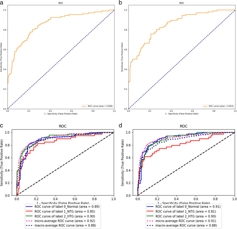
Methods: A cross-sectional, retrospective study in a tertiary referral hospital. Patients with healthy optic disc, high-tension, or normal-tension glaucoma were enrolled. Complicated non-glaucomatous optic neuropathy was excluded. Colour and red-free fundus images were collected for development of DLS and comparison of their efficacy. The convolutional neural network with the pre-trained EfficientNet-b0 model was selected for machine learning. Glaucoma screening (Binary) and ternary classification with or without additional demographics (age, gender, high myopia) were evaluated, followed by creating confusion matrix and heatmaps. Area under receiver operating characteristic curve (AUC), accuracy, sensitivity, specificity, and F1 score were viewed as main outcome measures.

Results: Two hundred and twenty-two cases (421 eyes) were enrolled, with 1851 images in total (1207 normal and 644 glaucomatous disc). Train set and test set were comprised of 1539 and 312 images, respectively. If demographics were not provided, AUC, accuracy, precision, sensitivity, F1 score, and specificity of our deep learning system in eye-based glaucoma screening were 0.98, 0.91, 0.86, 0.86, 0.86, and 0.94 in test set. Same outcome measures in eye-based ternary classification without demographic data were 0.94, 0.87, 0.87, 0.87, 0.87, and 0.94 in our test set, respectively. Adding demographics has no significant impact on efficacy, but establishing a linkage between eyes and images is helpful for a better performance. Confusion matrix and heatmaps suggested that retinal lesions and quality of photographs could affect classification. Colour fundus images play a major role in glaucoma classification, compared to red-free fundus images.

Conclusions: Promising results with high AUC and specificity were shown in distinguishing normal optic nerve from glaucomatous fundus images and doing further classification.

Keywords: Colour fundus photograph; Deep learning system; Glaucoma screening and classification; High myopia; Normal- tension glaucoma.


Conflict of interest statement

The authors declare that they have no competing interests.

Figures

Fig. 1
Fig. 1
The ROC curves with AUC in binary and ternary classification with or without demographics. Binary classification with (a) and without (b) information of age, gender, and high myopia in test set. Ternary classification with (c) and without (d) information of age, gender, and high myopia in test set
Fig. 2
Fig. 2
Image- or eye-based confusion matrix in test set of binary and ternary classification. Image- (a) and eye-based (b) analysis of binary classification (0.0 = normal; 1.0 = glaucoma) in test set. After adding information of age, gender, and high myopia, results of binary classification in test set with image- (c) or eye-based (d) analysis. Image- (e) and eye-based (f) analysis of ternary classification (0.0 = normal; 1.0 = normal-tension glaucoma; 2.0 = high-tension glaucoma) in test set. After adding information of age, gender, and high myopia, results of ternary classification in test set with image- (g) or eye-based (h) analysis
Fig. 3
Fig. 3
The outcome measures in image-based analysis of red-free or colour fundus images. Test metrics calculated from red-free fundus images and colour fundus images were compared by paired t-test. In binary classification, red-free fundus images achieved better performance in number, which was not statistically significant (a). Colour fundus images achieved better performance in ternary classification, in which statistically significant differences were observed (b). n.s. = not statistically significant
Fig. 4
Fig. 4
Binary classification presented by heatmap to show weighted area of deep learning system. Non-glaucomatous fundus (a) showed a weighted area peripherally, outside optic disc (b). Weighted area presented temporal to optic disc in normal-tension glaucoma (c and d) and high-tension glaucoma (e and f) in binary classification. Red-free fundal picture and its associated heatmap (g and h) in our study
Fig. 5
Fig. 5
Ternary classification presented by heatmap to show weighted area of deep learning system. Non-glaucomatous fundus (a) showed weighted area supero-temporal to optic disc (b). Weighted area presented at optic disc in high-tension glaucoma (c and d) in ternary classification. Misclassification of normal-tension glaucoma into high-tension glaucoma (e), showing weighted area nasal to optic disc (f) in the left eye. High-tension glaucoma was misclassified into normal-tension glaucoma (g), presenting a weighted area inferior to optic disc (h) in the right eye

References

    1. Quigley HA, Broman AT. The number of people with glaucoma worldwide in 2010 and 2020. Br J Ophthalmol. 2006;90:262–267. doi: 10.1136/bjo.2005.081224. - DOI - PMC - PubMed
    1. Shon K, Wollstein G, Schuman JS, Sung KR. Prediction of glaucomatous visual field progression: pointwise analysis. Curr Eye Res. 2014;39:705–710. doi: 10.3109/02713683.2013.867353. - DOI - PMC - PubMed
    1. Michelessi M, Lucenteforte E, Oddone F, Brazzelli M, Parravano M, Franchi S, et al. Optic nerve head and fiber layer imaging for diagnosing glaucoma. version 2. Cochrane Database Syst Rev. 2015;11:CD008803. - PMC - PubMed
    1. Sayres R, Taly A, Rahimy E, Blumer K, Coz D, Hammel N, et al. Using a deep learning algorithm and integrated gradients explanation to assist grading for diabetic retinopathy. Ophthalmology. 2019;126(4):552–64. doi: 10.1016/j.ophtha.2018.11.016. - DOI - PubMed
    1. Burlina PM, Joshi N, Pekala M, Pacheco KD, Freund DE, Bressler NM. Automated grading of age-related macular degeneration from color fundus images using deep convolutional neural networks. JAMA Ophthalmol. 2017;135:1170–1176. doi: 10.1001/jamaophthalmol.2017.3782. - DOI - PMC - PubMed