. 2023 Feb 14;11(1):e0387222.
doi: 10.1128/spectrum.03872-22. Epub 2022 Dec 13.

Three Amino Acid Substitutions in the Spike Protein Enable the Coronavirus Porcine Epidemic Diarrhea Virus To Infect Vero Cells

Affiliations

Three Amino Acid Substitutions in the Spike Protein Enable the Coronavirus Porcine Epidemic Diarrhea Virus To Infect Vero Cells

Bingqing Chen et al. Microbiol Spectr. .

Abstract

Porcine epidemic diarrhea virus (PEDV), a continuously evolving pathogen, causes severe diarrhea in piglets, with high mortality rates. To prevent or mitigate the disease, it is common practice to develop live or inactivated PEDV vaccines based on cell-adapted viral variants. Propagating wild-type PEDV in cultured cells is, however, often challenging due to the lack of knowledge about the requirements for the cell adaptation of PEDV. In the present study, by using the RNA-targeted reverse genetic system for PEDV to apply S protein swapping followed by the rescue of the recombinant viruses, three key amino acid mutations in the S protein, A605E, E633Q, and R891G, were identified, which enable attenuated PEDV strain DR13 (DR13att) to efficiently and productively infect Vero cells, in contrast to the parental DR13 strain (DR13par). The former two key mutations reside inside and in the vicinity of the receptor binding domain (RBD), respectively, while the latter occurs at the N-terminal end of the fusion peptide (FP). Besides the three key mutations, other mutations in the S protein further enhanced the infection efficiency of the recombinant viruses. We hypothesize that the three mutations changed PEDV tropism by altering the S2' cleavage site and the RBD structure. This study provides basic molecular insight into cell adaptation by PEDV, which is also relevant for vaccine design. IMPORTANCE Porcine epidemic diarrhea virus (PEDV) is a lethal pathogen for newborn piglets, and an efficient vaccine is needed urgently. However, propagating wild-type PEDV in cultured cells for vaccine development is still challenging due to the lack of knowledge about the mechanism of the cell adaptation of PEDV. In this study, we found that three amino acid mutations, A605E, E633Q, and R891G, in the spike protein of the Vero cell-adapted PEDV strain DR13att were critical for its cell adaptation. After analyzing the mutation sites in the spike protein, we hypothesize that the cell adaptation of DR13att was achieved by altering the S2' cleavage site and the RBD structure. This study provides new molecular insight into the mechanism of PEDV culture adaptation and new strategies for PEDV vaccine design.

Keywords: PEDV; S protein; cell adaptation; reverse genetics; tropism.


Conflict of interest statement

The authors declare no conflict of interest.

Figures
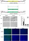
FIG 1
FIG 1
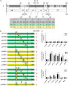
The S gene determines Vero cell adaptation of PEDV DR13att. The rescue of recombinant viruses (r-Spar and r-Satt) was carried out by using a reverse genetic system based on homologous RNA recombination. Briefly, LR7 cells infected with mPEDV (a recombinant PEDV with the DR13att backbone carrying the ectodomain of the S gene of mouse hepatitis coronavirus, thereby enabling the virus to infect murine LR7 cells) were harvested. Next, capped runoff RNA transcripts (donor RNA) synthesized from the PacI-linearized transfer vectors were transfected into the mPEDV-infected LR7 cells by electroporation. Finally, the electroporated cells were overlaid onto confluent Vero cells and cultured in the presence or absence of trypsin for the rescue of recombinant viruses, and candidate recombinant viruses were purified by two rounds of endpoint dilutions. (A) Construction of the transfer vector p-PEDV-Spar-ΔORF3/RLuc. The transfer vector contains the truncated 1a/1b and the structural genes of the PEDV genome. The thick red arrow indicates the T7 promoter from which donor RNA was synthesized in vitro using T7 RNA polymerase. The S gene of the PEDV DR13 parental strain (Spar) (GenBank accession number DQ862099) was obtained by using fusion PCR with artificially synthesized DNA fragments. p-PEDV-Spar-ΔORF3/RLuc was constructed by replacing the S gene of the PEDV DR13 attenuated strain (Satt) in p-PEDV-Satt-ΔORF3/RLuc with the Spar gene. UTR, untranslated region. (B) CPE of r-Spar and r-Satt on Vero cells. Vero cells in 24-well plates were infected with r-Spar (its culture) and r-Satt in the presence or absence of trypsin (15 μg/mL). The formation of CPE was observed at 36 h postinoculation (hpi). Arrows indicate CPE. Bar, 100 μm. (C) Luciferase expression by r-Spar and r-Satt. The intracellular Renilla luciferase activities of the first two generations (P0 and P1) of recombinant viruses were determined when obvious CPE appeared or at 5 days postinoculation (y axis) (relative light units [RLU]). The results are expressed as the mean values from three parallel tests, and error bars represent the standard deviations (SD). Comparisons were carried out between the luciferase activities of the recombinant viruses and that of the mock treatments. *, P < 0.05; **, P < 0.01. (D) Indirect immunofluorescence assay (IFA) of r-Spar- and r-Satt-infected cells. Vero cells were infected with r-Satt and r-Spar in the presence or absence of trypsin (15 μg/mL). Infected cells were fixed at 36 hpi and immunolabeled with rabbit anti-PEDV M polyclonal antibody (green). Nuclei were labeled with DAPI (blue).
FIG 2
FIG 2
The region of Satt from aa 530 to 590 contains a critical residue(s) determining the Vero cell adaptation of PEDV DR13att. (A) Schematic representation of the chimeric S proteins of the recombinant viruses r-Satt(1–529 aa)par, r-Satt(530–936 aa)par, r-Satt(937–1383 aa)par, and r-Spar(530–936 aa)att. (B) Luciferase expression by r-Satt(1–529 aa)par, r-Satt(530–936 aa)par, r-Satt(937–1383 aa)par, and r-Spar(530–936 aa)att. The intracellular Renilla luciferase activities of the first two generations (P0 and P1) of recombinant viruses were determined (y axis) (relative light units [RLU]). The results are expressed as the mean values from three parallel tests, and error bars represent the SD. Comparisons were made between the luciferase activities of the recombinant viruses and those of the mock treatments. *, P < 0.05; **, P < 0.01. (C) RT-PCR confirmation of the rescued recombinant PEDVs. RT-PCR was performed covering the region of the S protein from aa 530 to 936 (Primers Ped136 [5′-TGCATCTCGGTTTGTTGGATGC-3′] and Ped137 [5′-TATATTACCAATAGCAGAGTTA-3′]) using the RNA template isolated from the recombinant PEDVs, and the protein was analyzed by gel electrophoresis. The expected size of the RT-PCR product was 1,789 bp.
FIG 3
FIG 3
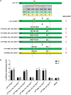
Mapping the exact region determining the Vero cell adaptation of DR13att. (A) Schematic representation of the Spar protein. the Spar protein is symbolically divided into S1 (aa 1 to 725) and S2 (aa 726 to 1383) subunits. The signal peptide (SP) (aa 1 to 18), regions containing a neutralizing epitope (COE) (aa 499 to 638), the fusion peptide (FP) (aa 891 to 908), two heptad repeat regions (HR1 [aa 978 to 1117] and HR2 [aa 1274 to 1313]), and the transmembrane domain (TM) (aa 1324 to 1346) are shown in light gray. The positions of the different amino acids are shown schematically as gray vertical lines below, and the numbers of amino acids differing between Satt and Spar in the corresponding regions are shown in parentheses. The region of Satt and Spar from aa 530 to 936 was divided into five groups (A to D), and the amino acid differences are listed in the table below. (B) Schematic representation of the chimeric S proteins. The positions of the mutated residues are shown, and their colors are consistent with the colors of their original S proteins. Dashed boxes refer to the groups defined as described above for panel A. (C) Luciferase expression by recombinant PEDVs with chimeric S genes. The intracellular Renilla luciferase activities of the first two generations of the recombinant viruses (P0 and P1) (y axis) (relative light units [RLU]) were determined. The results are expressed as the mean values from three parallel tests, and error bars represent the SD. Results of comparisons between the luciferase activities of the recombinant viruses and that of the mock treatments are indicated. *, P < 0.05; **, P < 0.01.
FIG 4
FIG 4
The combination of mutations at positions 603, 633, and 891 of Satt largely determines the Vero cell adaptation of DR13att. (A) Schematic representation of the chimeric S proteins of recombinant PEDVs. The positions of the mutated residues are shown, and their colors are consistent with the colors of their original S proteins. (B) Luciferase expression by recombinant PEDVs. The intracellular Renilla luciferase activities of the first two generations of the recombinant viruses (P0 and P1) (y axis) (relative light units [RLU]) were determined. The results are expressed as the mean values from three parallel tests, and error bars represent the SD. Comparisons were carried out between the luciferase activities of the recombinant viruses and that of the mock treatments. *, P < 0.05; **, P < 0.01.
FIG 5
FIG 5
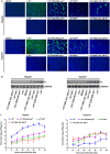
Characterization of the recombinant PEDVs in Vero cells. (A) IFA of recombinant PEDVs. Eight recombinant PEDVs, r-Spar, r-Satt, r-Satt(530–936 aa)par, r-Spar(530–936 aa)att, r-SattAEpar, r-SparAEatt, r-Satt(605, 633, 891)par, and r-Spar(605, 633, 891)att, were inoculated into Vero cells in the presence or absence of trypsin (15 μg/mL). Cells were fixed at 36 hpi and immunolabeled with rabbit anti-PEDV M polyclonal antibody (green). Nuclei were labeled with DAPI (blue). (B) Western blot analysis of Vero cells infected with recombinant PEDVs. Vero cells inoculated with recombinant PEDVs were harvested at 24 hpi. Proteins in the lysed cells were separated by 12% SDS-PAGE and subsequently processed for immunoblot analysis with monoclonal antibody against the PEDV N protein; GAPDH served as a protein loading control. (C) Multistep growth kinetics of recombinant PEDVs. Vero cells were inoculated with r-Satt, r-Spar(530–936 aa)att, r-SparAEatt, and r-Spar(605, 633, 891)att (MOI of 0.0012) and continued to be cultured in the presence or absence of trypsin (15 μg/mL). At the indicated times postinfection, cells and culture media were harvested by three rounds of freezing and thawing, followed by centrifugation to remove cell debris. The supernatants were collected and used to measure viral titers in the presence of trypsin. Results are expressed as the mean values from three parallel tests, and error bars represent the SD.

Similar articles

Cited by

References

    1. Lee C. 2015. Porcine epidemic diarrhea virus: an emerging and re-emerging epizootic swine virus. Virol J 12:193. doi:10.1186/s12985-015-0421-2. - DOI - PMC - PubMed
    1. Jung K, Saif LJ. 2015. Porcine epidemic diarrhea virus infection: etiology, epidemiology, pathogenesis and immunoprophylaxis. Vet J 204:134–143. doi:10.1016/j.tvjl.2015.02.017. - DOI - PMC - PubMed
    1. Sun J, Li Q, Shao C, Ma Y, He H, Jiang S, Zhou Y, Wu Y, Ba S, Shi L, Fang W, Wang X, Song H. 2018. Isolation and characterization of Chinese porcine epidemic diarrhea virus with novel mutations and deletions in the S gene. Vet Microbiol 221:81–89. doi:10.1016/j.vetmic.2018.05.021. - DOI - PMC - PubMed
    1. Chen N, Li S, Zhou R, Zhu M, He S, Ye M, Huang Y, Li S, Zhu C, Xia P, Zhu J. 2017. Two novel porcine epidemic diarrhea virus (PEDV) recombinants from a natural recombinant and distinct subtypes of PEDV variants. Virus Res 242:90–95. doi:10.1016/j.virusres.2017.09.013. - DOI - PubMed
    1. Sun R-Q, Cai R-J, Chen Y-Q, Liang P-S, Chen D-K, Song C-X. 2012. Outbreak of porcine epidemic diarrhea in suckling piglets, China. Emerg Infect Dis 18:161–163. doi:10.3201/eid1801.111259. - DOI - PMC - PubMed

Publication types

Substances

LinkOut - more resources