. 2023 Jun;33(6):3867-3877.
doi: 10.1007/s00330-022-09325-x. Epub 2022 Dec 13.

Cardiac MRI in patients with COVID-19 infection

Affiliations

Cardiac MRI in patients with COVID-19 infection

Emad H Abdeldayem et al. Eur Radiol. 2023 Jun.

Abstract

Objective: COVID-19 infection is a systemic disease with various cardiovascular symptoms and complications. Cardiac MRI with late gadolinium enhancement is the modality of choice for the assessment of myocardial involvement. T1 and T2 mapping can increase diagnostic accuracy and improve further management. Our study aimed to evaluate the different aspects of myocardial damage in cases of COVID-19 infection using cardiac MRI.

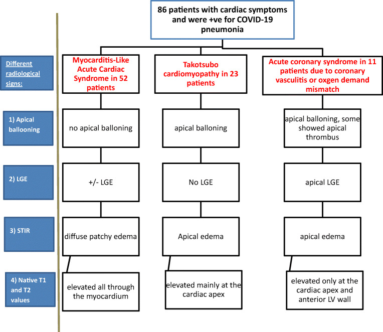
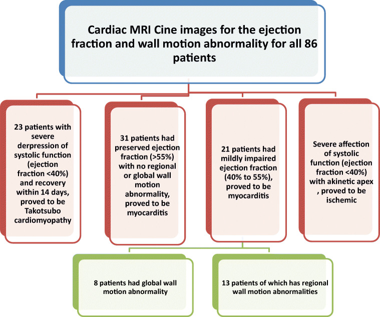
Methods: This descriptive retrospective study included 86 cases, with a history of COVID-19 infection confirmed by positive RT-PCR, who met the inclusion criteria. Patients had progressive chest pain or dyspnoea with a suspected underlying cardiac cause, either by an abnormal electrocardiogram or elevated troponin levels. Cardiac MRI was performed with late contrast-enhanced (LGE) imaging, followed by T1 and T2 mapping.

Results: Twenty-four patients have elevated hsTnT with a median hsTnT value of 133 ng/L (IQR: 102 to 159 ng/L); normal value < 14 ng/L. Other sixty-two patients showed elevated hsTnI with a median hsTnI value of 1637 ng/L (IQR: 1340 to 2540 ng/L); normal value < 40 ng/L. CMR showed 52 patients with acute myocarditis, 23 with Takotsubo cardiomyopathy, and 11 with myocardial infarction. Invasive coronary angiography was performed only in selected patients.

Conclusion: Different COVID-19-related cardiac injuries may cause similar clinical symptoms. Cardiac MRI is the modality of choice to differentiate between the different types of myocardial injury such as Takotsubo cardiomyopathy and infection-related cardiomyopathy or even acute coronary syndrome secondary to vasculitis or oxygen-demand mismatch.

Key points: • It is essential to detect early COVID-related cardiac injury using different cardiac biomarkers and cardiac imaging, as it has a significant impact on patient management and outcome. • Cardiac MRI is the modality of choice to differentiate between the different aspects of COVID-related myocardial injury.

Keywords: Acute coronary syndrome; Coronavirus; Magnetic resonance imaging; Myocarditis; Takotsubo cardiomyopathy.


Conflict of interest statement

The authors of this manuscript declare no relationships with any companies whose products or services may be related to the subject matter of the article.

Figures

Fig. 1
Fig. 1
Detection of the different radiological signs using the cardiac MRI sequences for characterization of the cardiac complications in patients with COVID-19 infection
Fig. 2
Fig. 2
Algorithm representing the spectrum of analysis of cardiac MRI finding regarding the end-diastolic volume and approach through the abnormal finding to differentiate between cardiac complications in patients with COVID-19 infection
Fig. 3
Fig. 3
Algorithm representing the spectrum of analysis of cardiac MRI finding regarding the ejection fraction and wall motion abnormality to differentiate between cardiac complications in patients with COVID-19 infection
Fig. 4
Fig. 4
Cardiac MRI: a white blood image 2-chamber vertical long-axis view showing ballooning of the cardiac apex. b T2-weighted-STIR image 2-chamber view showing interstitial edema of the cardiac apex. c IR post-contrast image 4-chamber view showing no appreciable LGE of the cardiac apex. d, e T2 and T1 cardiac MRI mapping sequence in two-chamber views respectively showing intense interstitial myocardial edema (native T2 = 64 ms) and elevated T1 value =1178 ms. Conventional coronary angiography of LAD confirmed normal appearance with no stenosis or occlusion. The final diagnosis was Takotsubo-like cardiomyopathy (as a part of acute myocarditis COVID-like syndrome (AMCovS)
Fig. 5
Fig. 5
Cardiac MRI: a white blood image 2-chamber short-axis view showing preserved shape and configuration of the left ventricle. b T2-weighted-STIR image 2-chamber short axis showing faint patchy interstitial edema of the myocardium. c IR post-contrast image 2-chamber view showing multiple patchy mid-myocardial enhancing zones of the late gadolinium enhancement, more evident at basal septal and inferior walls. d, e T2 and T1 cardiac MRI mapping sequence in two-chamber view respectively showing intense interstitial myocardial edema with native T2 = 67 ms and elevated T1 value = 1154 ms. Conventional coronary angiography of LAD confirmed normal appearance with no stenosis or occlusion. The final diagnosis was acute myocarditis COVID-like syndrome (AMCovS)
Fig. 6
Fig. 6
Cardiac MRI: a White blood image 4-chamber view showing ballooning and thinning out of the cardiac apex with apical thrombus. b T2-weighted-STIR image 4-chamber view showing faint interstitial edema of the cardiac apex and apical cardiac segments. c IR post-contrast image 4-chamber view showing sub-endocardial / trans-mural enhancing zones of the late gadolinium enhancement along with the LAD territory. d, e T2 and T1 cardiac MRI mapping sequence in two-chamber view respectively showing mild interstitial myocardial edema (native T2 = 58 ms) and elevated T1 value = 1193 ms. Conventional coronary angiography of LAD confirmed tapering and narrowing of distal LAD. The LCX and RCA were normal. The final diagnosis was acute coronary syndrome due to coronary vasculitis or oxygen demand mismatch

References

    1. Vaninov N. In the eye of the COVID-19 cytokine storm. Nat Rev Immunol. 2020;20:277. doi: 10.1038/s41577-020-0305-6. - DOI - PMC - PubMed
    1. Esfandiarei M, McManus BM. Molecular biology and pathogenesis of viral myocarditis. Annu Rev Pathol. 2008;3:127–155. doi: 10.1146/annurev.pathmechdis.3.121806.151534. - DOI - PubMed
    1. Caforio AL, Pankuweit S, Arbustini E, et al. Current state of knowledge on etiology, diagnosis, management, and therapy of myocarditis: a position statement of the European Society of Cardiology Working Group on Myocardial and Pericardial Diseases. Eur Heart J. 2013;34:2636–2648. doi: 10.1093/eurheartj/eht210. - DOI - PubMed
    1. Kociol RD, Cooper LT, Fang JC, et al. Recognition and initial management of fulminant myocarditis: a scientific statement from the American Heart Association. Circulation. 2020;141:e69–e92. doi: 10.1161/CIR.0000000000000745. - DOI - PubMed
    1. Siripanthong B, Nazarian S, Muser D, et al. Recognizing COVID-19–related myocarditis: the possible pathophysiology and proposed guideline for diagnosis and management. Heart Rhythm. 2020;17:1463–1471. doi: 10.1016/j.hrthm.2020.05.001. - DOI - PMC - PubMed