

LATE-NC staging in routine neuropathologic diagnosis: an update
- PMID: 36512061
- PMCID: PMC9849315
- DOI: 10.1007/s00401-022-02524-2
LATE-NC staging in routine neuropathologic diagnosis: an update
Abstract
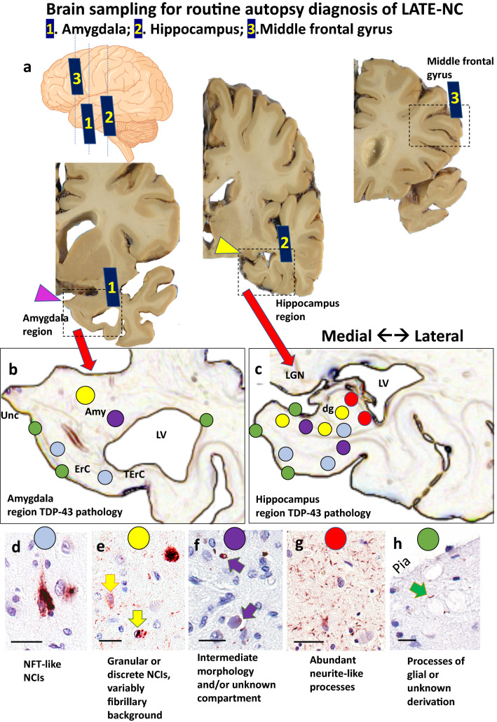
An international consensus report in 2019 recommended a classification system for limbic-predominant age-related TDP-43 encephalopathy neuropathologic changes (LATE-NC). The suggested neuropathologic staging system and nomenclature have proven useful for autopsy practice and dementia research. However, some issues remain unresolved, such as cases with unusual features that do not fit with current diagnostic categories. The goal of this report is to update the neuropathologic criteria for the diagnosis and staging of LATE-NC, based primarily on published data. We provide practical suggestions about how to integrate available genetic information and comorbid pathologies [e.g., Alzheimer's disease neuropathologic changes (ADNC) and Lewy body disease]. We also describe recent research findings that have enabled more precise guidance on how to differentiate LATE-NC from other subtypes of TDP-43 pathology [e.g., frontotemporal lobar degeneration (FTLD) and amyotrophic lateral sclerosis (ALS)], and how to render diagnoses in unusual situations in which TDP-43 pathology does not follow the staging scheme proposed in 2019. Specific recommendations are also made on when not to apply this diagnostic term based on current knowledge. Neuroanatomical regions of interest in LATE-NC are described in detail and the implications for TDP-43 immunohistochemical results are specified more precisely. We also highlight questions that remain unresolved and areas needing additional study. In summary, the current work lays out a number of recommendations to improve the precision of LATE-NC staging based on published reports and diagnostic experience.
Keywords: Aging; Dementia; FTD; Hippocampal sclerosis; NCI; Neuroanatomy; Processes; Stages; TDP-43.
© 2022. The Author(s).
Conflict of interest statement
E.B.L., D.W.D., M.N., D.R.T., G.H., G.G.K., and P.T.N. are members of the Editorial Board of
Figures
References
-
- (2022) LATE 2022 Webinar, 11 February https://www.nia.nih.gov/research/dn/late-2022. Accessed 1 Nov 2022
-
- Agirre-Beitia G, Moreno-Estebanez A, Gonzalez-Pinto Gonzalez T, Melgar Jimenez B, Campos Rodriguez I, Cabral Martinez L, et al. FOSMN: a possible TDP-43 proteinopathy to consider in a patient with facial sensory symptoms. Neurol Clin Pract. 2020;10:e47–e50. doi: 10.1212/CPJ.0000000000000753. - DOI - PMC - PubMed
-
- Agrawal S, Leurgans SE, James BD, Barnes LL, Mehta RI, Dams-O'Connor K, et al. Association of traumatic brain injury with and without loss of consciousness with neuropathologic outcomes in community-dwelling older persons. JAMA Netw Open. 2022;5:e229311. doi: 10.1001/jamanetworkopen.2022.9311. - DOI - PMC - PubMed
-
- Agrawal S, Yu L, Nag S, Arfanakis K, Barnes LL, Bennett DA, et al. The association of Lewy bodies with limbic-predominant age-related TDP-43 encephalopathy neuropathologic changes and their role in cognition and Alzheimer’s dementia in older persons. Acta Neuropathol Commun. 2021;9:156. doi: 10.1186/s40478-021-01260-0. - DOI - PMC - PubMed
-
- Alafuzoff I, Pikkarainen M, Neumann M, Arzberger T, Al-Sarraj S, Bodi I, et al. Neuropathological assessments of the pathology in frontotemporal lobar degeneration with TDP43-positive inclusions: an inter-laboratory study by the BrainNet Europe consortium. J Neural Transm (Vienna) 2015;122:957–972. doi: 10.1007/s00702-014-1304-1. - DOI - PubMed
Publication types
MeSH terms
Substances
Supplementary concepts
Grants and funding
- G0900582/MRC_/Medical Research Council/United Kingdom
- K08 AG065463/AG/NIA NIH HHS/United States
- R01 AG061111/AG/NIA NIH HHS/United States
- P30 AG066512/AG/NIA NIH HHS/United States
- K24 AG053435/AG/NIA NIH HHS/United States
- R01 AG054449/AG/NIA NIH HHS/United States
- P30 AG062429/AG/NIA NIH HHS/United States
- G0601022/MRC_/Medical Research Council/United Kingdom
- P01 AG060882/AG/NIA NIH HHS/United States
- R01 AG064233/AG/NIA NIH HHS/United States
- P30 AG066509/AG/NIA NIH HHS/United States
- P30 AG072976/AG/NIA NIH HHS/United States
- P30 AG072972/AG/NIA NIH HHS/United States
- G9901400/MRC_/Medical Research Council/United Kingdom
- P30 AG072979/AG/NIA NIH HHS/United States
- RF1 AG069052/AG/NIA NIH HHS/United States
- P30 AG066507/AG/NIA NIH HHS/United States
- P30 AG072946/AG/NIA NIH HHS/United States
- P30 AG010133/AG/NIA NIH HHS/United States
- U24 AG021886/AG/NIA NIH HHS/United States
- R01 AG067482/AG/NIA NIH HHS/United States
- UL1 TR002538/TR/NCATS NIH HHS/United States
- RF1 NS118584/NS/NINDS NIH HHS/United States
- R01 AG062706/AG/NIA NIH HHS/United States
- P30 AG066530/AG/NIA NIH HHS/United States
- P30 AG072977/AG/NIA NIH HHS/United States
- U19 AG062418/AG/NIA NIH HHS/United States
- K08 AG065426/AG/NIA NIH HHS/United States
LinkOut - more resources
Full Text Sources
Medical
Miscellaneous
