Review
. 2023 Jan;20(1):83-96.
doi: 10.1007/s13311-022-01332-6. Epub 2022 Dec 13.

Overexpression-Induced α-Synuclein Brain Spreading

Affiliations
Review

Overexpression-Induced α-Synuclein Brain Spreading

Rita Pinto-Costa et al. Neurotherapeutics. 2023 Jan.

Abstract

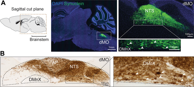
Interneuronal transfer of pathological α-synuclein species is thought to play an important role in the progressive advancement of Lewy pathology and increasing severity of clinical manifestations in Parkinson's and other diseases commonly referred to as synucleinopathies. Pathophysiological conditions and mechanisms triggering this trans-synaptic spreading bear therefore significant pathogenetic implications but have yet to be fully elucidated. In vivo experimental models support the conclusion that increased expression of intraneuronal α-synuclein can itself induce protein spreading throughout the brain as well as from the brain to peripheral tissues. For example, overexpression of α-synuclein targeted to the rodent dorsal medulla oblongata results in its transfer and accumulation into recipient axons innervating this brain region; through these axons, α-synuclein can then travel caudo-rostrally and reach other brain sites in the pons, midbrain, and forebrain. When protein overexpression is induced in the rodent midbrain, long-distance α-synuclein spreading can be followed over time; spreading-induced α-synuclein accumulation affects lower brain regions, including the dorsal motor nucleus of the vagus, proceeds through efferent axons of the vagus nerve, and is ultimately detected within vagal motor nerve endings in the gastric wall. As discussed in this review, animal models featuring α-synuclein overexpression not only support a relationship between α-synuclein burden and protein spreading but have also provided important clues on conditions/mechanisms capable of promoting interneuronal α-synuclein transfer. Intriguing findings include the relationship between neuronal activity and protein spreading and the role of oxidant stress in trans-synaptic α-synuclein mobility.

Keywords: Animal models; Gut-brain axis; Neuronal activity; Oxidative stress; Parkinson; Vagus nerve.


Figures

Fig. 1
Fig. 1
Targeted overexpression of hα-synuclein in the dorsal medulla oblongata. Images were obtained from mice that received an intravagal injection of hα-synuclein-AAVs and were sacrificed 2 weeks later. A The fluorescent image on the left shows hα-synuclein-containing neurons (green) in the dorsal medulla oblongata (dMO) in a sagittal brain section that was stained with both anti-hα-synuclein and DAPI (blue). The square rectangle delineates an area containing the nucleus of the tractus solitarius (NTS) and the dorsal motor nucleus of the vagus (DMnX) that is shown on the right top panel. The lower panel on the right shows transduced DMnX cell bodies at higher magnification (arrowheads). B Bright-field images show hα-synuclein-immunoreactive neurons (brown) in the DMnX, NTS and area postrema (AP) in a coronal section of the medulla oblongata. Arrowheads indicate transduced DMnX cell bodies in the high-magnification image
Fig. 2
Fig. 2
Neuronal accumulation of hα-synuclein as a result of caudo-rostral protein spreading. Images were obtained from rats that were injected intravagally with hα-synuclein-AAVs and sacrificed 6 weeks later. Coronal brain tissue sections were stained with anti-hα-synuclein. Bright-field images show immunoreactive axons (brown) with robustly labeled varicosities in the pons, hypothalamus, and thalamus
Fig. 3
Fig. 3
Schematic representation of long-distance brain-to-stomach hα-synuclein spreading. This spreading was initiated by an injection of hα-synuclein-AAVs in the rat midbrain (A). The exogenous protein first reached the dorsal medulla oblongata (dMO, box delineated by dashed lines) and, in particular, the dorsal motor nucleus of the vagus (B). It then traveled through efferent axons of the vagus nerve (C) and was finally detected within vagal nerve endings in the gastric wall (D)

References

    1. Braak H, Del Tredici K, Rüb U, De Vos RA, Steur ENJ, Braak E. Staging of brain pathology related to sporadic Parkinson’s disease. Neurobiol Aging. 2003;24(2):197–211. doi: 10.1016/S0197-4580(02)00065-9. - DOI - PubMed
    1. Braak H, Rüb U, Gai W, Del Tredici K. Idiopathic Parkinson's disease: possible routes by which vulnerable neuronal types may be subject to neuroinvasion by an unknown pathogen. J Neural Transm. 2003;110(5):517–536. doi: 10.1007/s00702-002-0808-2. - DOI - PubMed
    1. McCann H, Cartwright H, Halliday GM. Neuropathology of α-synuclein propagation and braak hypothesis. Mov Disord. 2016;31(2):152–160. doi: 10.1002/mds.26421. - DOI - PubMed
    1. Peng C, Trojanowski JQ, Lee VM-Y. Protein transmission in neurodegenerative disease. Nat Rev Neurol. 2020;16(4):199–212. doi: 10.1038/s41582-020-0333-7. - DOI - PMC - PubMed
    1. Breen DP, Halliday GM, Lang AE. Gut–brain axis and the spread of α-synuclein pathology: vagal highway or dead end? Mov Disord. 2019;34(3):307–316. doi: 10.1002/mds.27556. - DOI - PubMed

Publication types

Substances