. 2022 Dec 12;40(12):1470-1487.e7.
doi: 10.1016/j.ccell.2022.11.006.

Expression of inducible factors reprograms CAR-T cells for enhanced function and safety

Affiliations

Expression of inducible factors reprograms CAR-T cells for enhanced function and safety

Anže Smole et al. Cancer Cell. .

Abstract

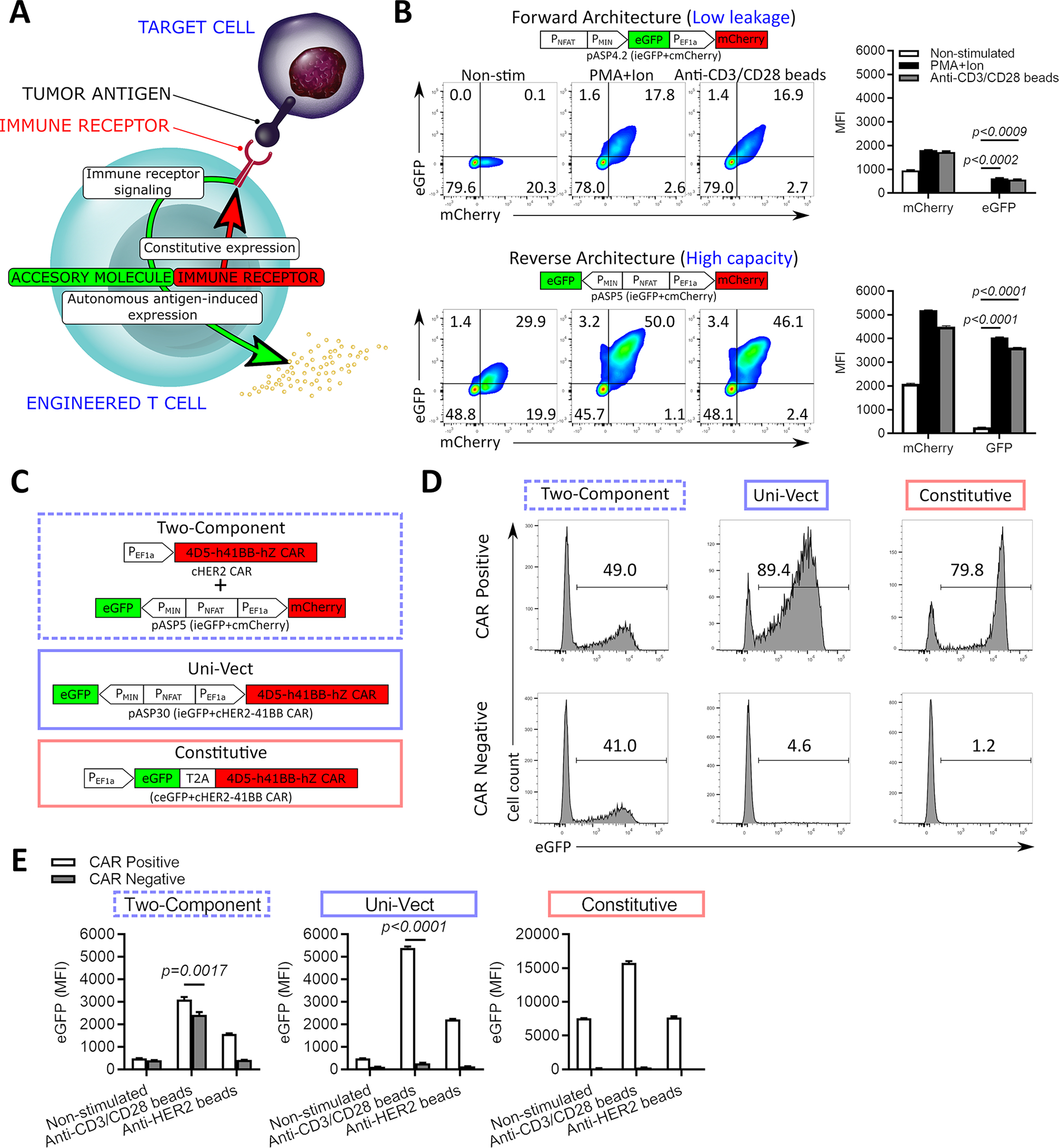
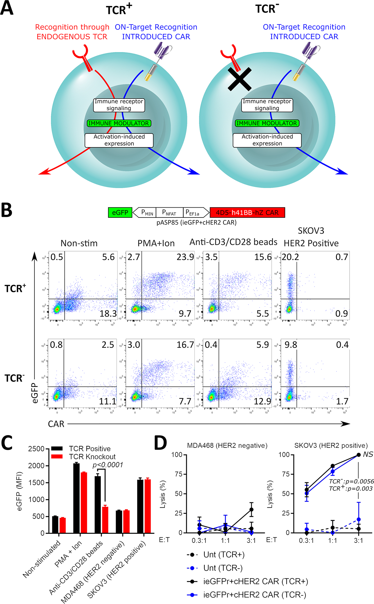
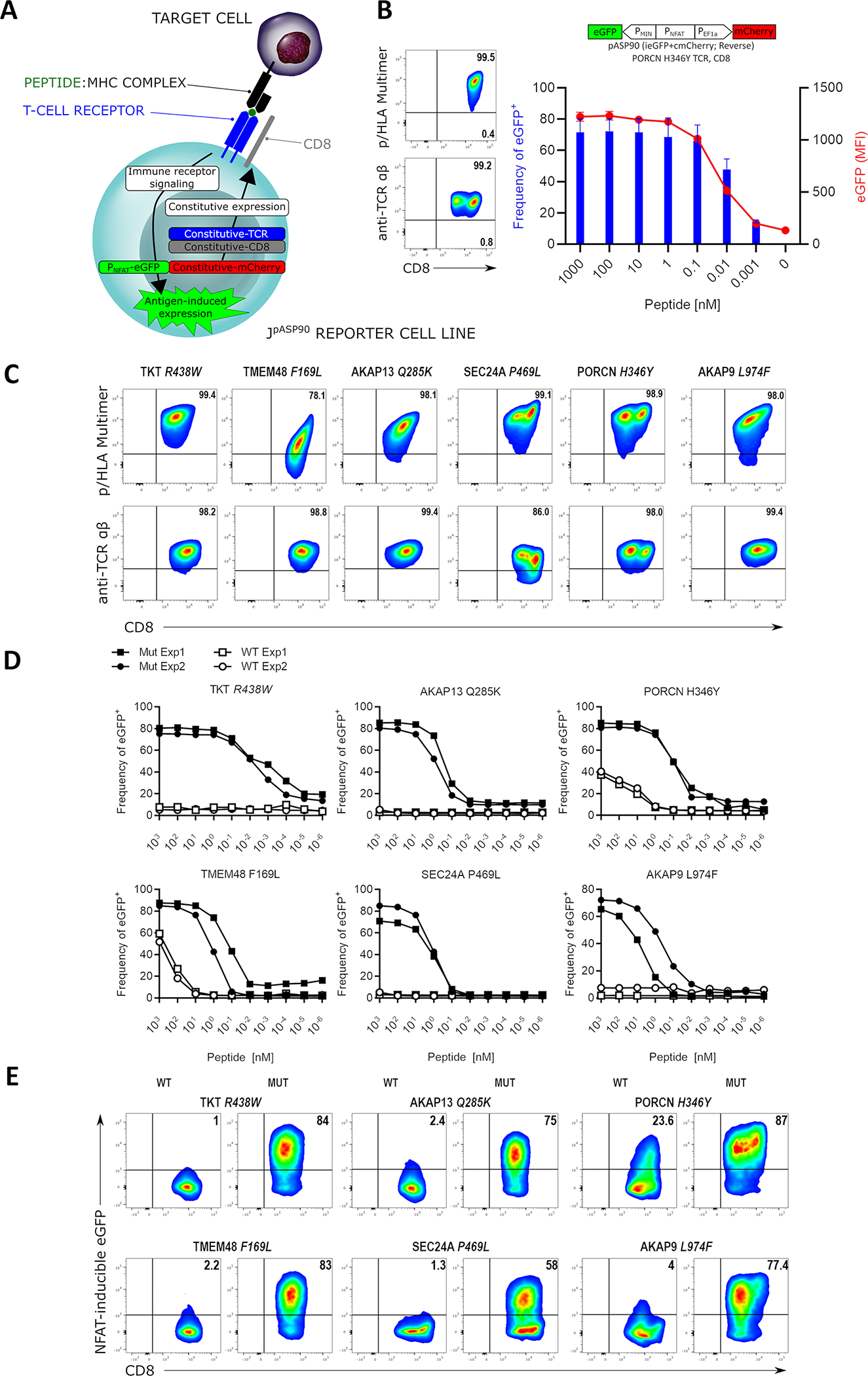
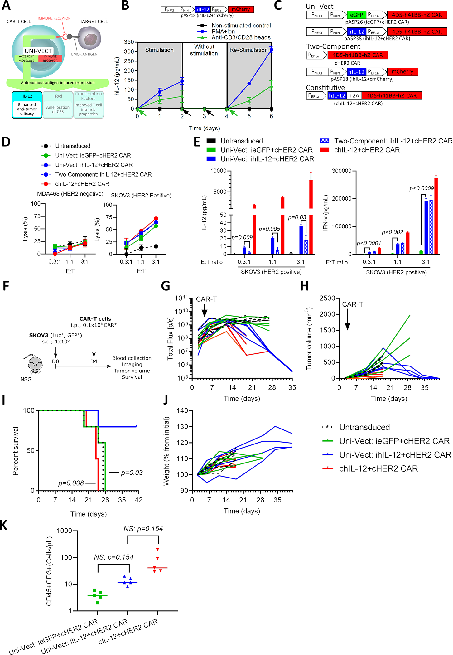
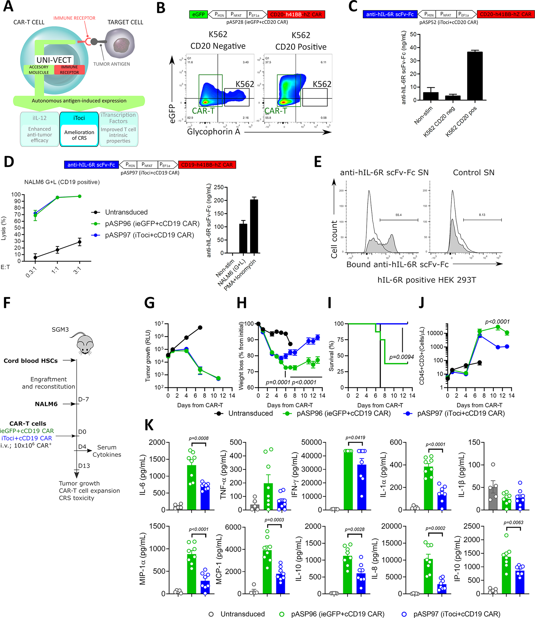
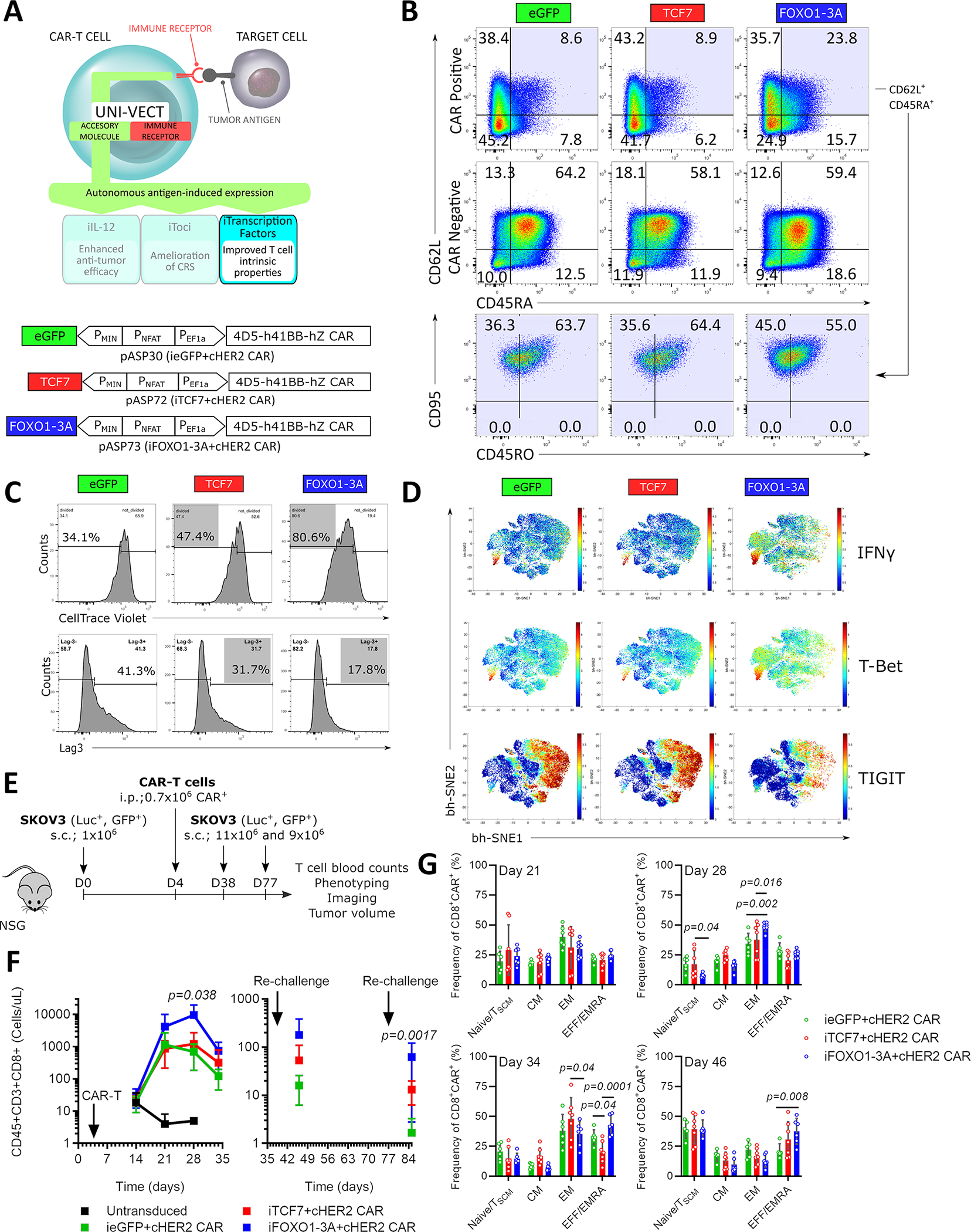
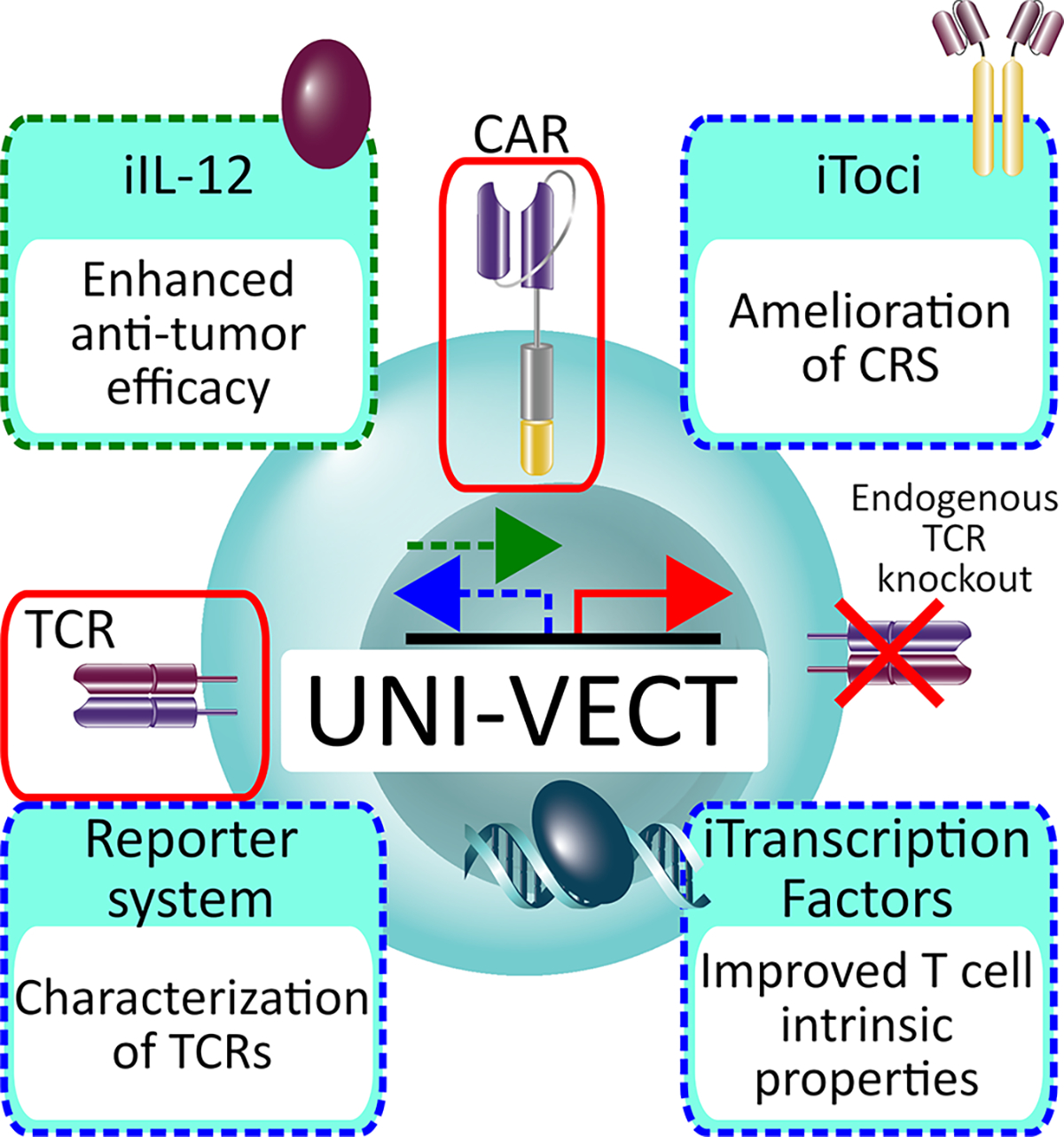
Despite the success of CAR-T cell cancer immunotherapy, challenges in efficacy and safety remain. Investigators have begun to enhance CAR-T cells with the expression of accessory molecules to address these challenges. Current systems rely on constitutive transgene expression or multiple viral vectors, resulting in unregulated response and product heterogeneity. Here, we develop a genetic platform that combines autonomous antigen-induced production of an accessory molecule with constitutive CAR expression in a single lentiviral vector called Uni-Vect. The broad therapeutic application of Uni-Vect is demonstrated in vivo by activation-dependent expression of (1) an immunostimulatory cytokine that improves efficacy, (2) an antibody that ameliorates cytokine-release syndrome, and (3) transcription factors that modulate T cell biology. Uni-Vect is also implemented as a platform to characterize immune receptors. Overall, we demonstrate that Uni-Vect provides a foundation for a more clinically actionable next-generation cellular immunotherapy.

Keywords: CAR-T Cells; CRS; IL-12; IL-6; NFAT; TCR; armored; inducible; single lentiviral expression system; transcription factor.


Conflict of interest statement

Declaration of interests A.S., A.D.P., C.H.J., and D.J.P. are co-inventors on PCT International Patent Applications by The Trustees of the University of Pennsylvania, which incorporate discoveries and inventions described here. Tmunity Therapeutics has licensed non-exclusive rights to commercialize licensed cytokine-secreting products, such as CAR- or TCR-modified T cells engineered to express a transgene-encoded cytokine. C.H.J. is a co-scientific founder of Tmunity Therapeutics and Capstan Therapeutics and is a member of the scientific advisory boards of Alaunos, BluesphereBio, Cabaletta, Carisma, Cellares, Poseida, Verismo, and viTToria bio. C.H.J. is an inventor on patents and/or patent applications licensed to Novartis Institutes of Biomedical Research and Tmunity Therapeutics and receives license revenue from such licenses. D.J.P. is a member of the scientific advisory boards for Bellicium Pharmaceuticals and InstilBio. D.J.P. is inventor on patents and/or patent applications licensed to Prescient Therapeutics, Tmunity Therapeutics, and Miltenyi Biotec and receives license revenue from those licenses.

Figures

Figure 1:
Figure 1:. Design and Validation of Uni-Vect in Primary Human T Cells
(A) Schematic representation of Uni-Vect recognizing a target antigen. (B) Primary human T cells were transduced with pASP4.2 or pASP5 at MOI5 and stimulated. Expression of reporter genes was monitored and quantified as MFI after 24 h. (n=3). (Welch ANOVA) (C) Schematic representation of compared constructs/experimental groups. (D-E) Primary human T cells were transduced with the vectors shown in C. Cells were sorted into CAR+ and CAR fractions then stimulated. (D) eGFP expression of cells activated with PMA+ionomycin overnight. Histograms are shown for CAR+ and CAR fractions. (E) Cells were left unstimulated or stimulated through TCRs or CARs for 24 h. MFI of eGFP is shown. (n=3). (Student’s T test). MFI; Mean fluorescence intensity. NS; not significant. All data with error bars are presented as mean ± SD. All data are representative of two or more experiments. See also Figures S1 and S2 and Table S1.
Figure 2:
Figure 2:. T-Cell Receptor Knockout in Uni-Vect CAR-T cells
(A) Uni-Vect-CAR-T cells may be stimulated by either the introduced CAR or the endogenous TCR. Endogenous TCR knockout prevents expression of the accessory molecule induced by endogenous TCR signaling. (B) TCR knockout in pASP85-CAR-T cells. NFAT signaling induced by 24 h stimulation with PMA+ionomycin, anti-CD3/CD28 dynabeads, or target SKOV3 cells was monitored in TCR+ and TCR CAR-T cells by flow cytometry via built-in NFAT-eGFP inducible module. (C) MFI quantification of NFAT-inducible eGFP in Q1 and Q2. (n=3) (Student’s T test) (D) Lysis of target and non-target cells by TCR+ and TCR pASP85-CAR-T cells (n=3). (ANOVA multiple-comparison). NS; not significant. All data with error bars are presented as mean ± SD. All data are representative of three or more experiments. See also Figure S3 and Table S1.
Figure 3:
Figure 3:. Uni-Vect Expression Cassettes for Characterization of T-cell Receptors
(A) Schematic representation of Uni-Vect-Jurkat reporter cell lines JpASP90 and JpASP89. (B) Transduction of TCR constructs into JpASP90 reporter cell lines. Titrated concentrations of peptide (LLHGFSFYL) antigen in the presence of antigen presenting cells led to upregulation of NFAT-inducible eGFP expression as a surrogate for activation via TCR signaling. Blue bars represent percentage of eGFP positive cells, red line represents MFI of eGFP at each concentration. (n=3). (C) Flow cytometry analysis of 6 neoantigen-specific/HLA-A*02:01 restricted TCRs engineered into the JpASP90 reporter cell line. (D) NFAT reporter EC50 curves for neoantigen-specific TCRs expressed in JpASP90 reporter cell line upon activation with mutated (blue) and wild-type (red) peptides. (E) JpASP90 reporter cell line expressing the various TCRs were cultured for 16 h with melanoma cell line DM6 expressing tandem mini-gene constructs encoding mutated or wild-type antigen and analyzed for inducible eGFP expression. All data are representative of two or more experiments. All data with error bars are presented as mean ± SD. See also Figure S4 and Tables S1 and S2.
Figure 4:
Figure 4:. iIL-12 Enabled Uni-Vect Improves Antitumor Efficacy of CAR-T Cells In Vivo
(A) Schematics of iIL-12 integrated into Uni-Vect. (B) Primary T cells transduced with pASP18 were stimulated for 2 days, followed by 2 days of rest, and then re-stimulated for another 2 days. IL-12 secretion was measured at 24 h increments. (n=3). Green arrow represents stimulation and black arrow represents media exchange. (C) Schematic representation of compared constructs/experimental groups. (D) CAR-T cells were co-cultured with HER2+ and HER2 target cell lines. Lysis was measured by luciferase assay. (n=3) (E) IL-12 and IFN-γ secretion from co-cultures in (D). (Welch ANOVA) (F) In vivo study design. CAR-T cells were injected i.p. at 0.1 × 10⁶ CAR-T per mouse in NSG mice with established HER2+ SKOV3 ovarian cancer tumors. (n = 5 mice per group) (G) Quantified tumor luciferase activity. (H) Tumor volume as measured by caliper. (I) Percent survival in each group (Log-rank Mantel-Cox test). (J) Plot of mouse weight versus time by group (n=5). (K) CD3+ T cell counts in peripheral blood at day 17. (Kruskal-Wallis test). i.p; Intraperitoneal, s.c.; Subcutaneous. Each line in G, H, and J and each dot in K represents individual mice. NS; not significant. All data are representative of two or more experiments. All data with error bars are presented as mean ± SD. See also Figure S5 and Table S1.
Figure 5:
Figure 5:. Antigen-inducible Secretion of Biologically Active Tocilizumab-based scFv-Fc from CAR-T cells
(A) Schematics of iToci integrated into Uni-Vect. (B) pASP28-CAR-T cells were co-cultured with CD20+ or CD20 K562 cells at 5:1 effector to target ratio for 24 h. Flow graph represents inducible eGFP, concomitant with target cell lysis. K562 cell lines (black rectangle) were stained for Glycophorin A expression and appear as Glycophorin+eGFP population. CAR-T cells appear in Glycophorin population (green rectangle). (C) pASP52-CAR-T cells were co-cultured with CD20+ or CD20 K562 cells at 3:1 effector to target ratio for 72 h. Antigen-dependent secretion of iToci was measured. (n=3) (D) pASP97-CAR-T cells or control pASP96-CAR-T cells were co-cultured with CD19+ NALM6 cell line for 48 h. Lysis was measured by luciferase assay (Left) and concomitant secretion of iToci was measured by ELISA (Right). (n=3) (E) pASP52-CAR-T cells or control pASP28-CAR-T cells were co-cultured with CD20+ cell line. Supernatants from these co-cultures were added to the HEK 293T cells engineered to express hIL-6Rα and binding of iToci was examined. (F) In vivo study design. pASP97-CAR-T cells or control pASP96-CAR-T cells were injected in humanized SGM3 mice engrafted with CD19+ NALM6 cell line. (n = 8 mice per group for iToci+cCD19 CAR and ieGFP+cCD19 CAR; n = 5 for untransduced group) Experiment was performed once. We monitored (G) tumor growth control, (H) weight loss (Two-Way ANOVA), (I) survival of mice (Log-rank Mantel-Cox test) and (J) hCD45+CD3+ T cell numbers in peripheral blood (Two-Way ANOVA). (K) Inflammatory cytokines were measured in serum 4 days after CAR-T cells injection (Two-Way ANOVA). SN, supernatant. NS; not significant. Error bars are presented as mean ± SD in C and D and as mean ± SEM in G, H, J and K. See also Figure S6 and Table S1.
Figure 6:
Figure 6:. Inducible Transcription Factors Program Therapeutically Relevant CAR-T Cell States and Improve Expansion In Vivo
(A) Schematics of iTFs integrated into Uni-Vect; pASP30, pASP72, and pASP73 Uni-Vect constructs. (B) iTF-CAR-T cells were exposed to SKOV3 cells twice (Detailed in Figure S7A). Phenotype was analyzed 3 days after second stimulation. Data is representative of 2 experiments performed. (C) iTF-CAR-T cells were sorted for CAR+ population and exposed to repeated stimulations with SKOV3 cells (Detailed in Figure S7A). Upper: After 2nd stimulation, iTF-CAR-T cells were labeled with CellTrace Violet and re-exposed to SKOV3 cells for 5 days. Proliferation was measured. Lower: Lag3 expression after 3rd stimulation. (D) 3 days after 4th round of stimulation described in (C), T cell phenotype markers were analyzed by CyTOF. (E) In vivo study design. n = 7 mice per experimental group. After initial tumor clearance, mice were re-challenged twice with SKOV3. (F) CD8+ T cell counts in peripheral blood. Left: initial response and Right: after re-challenges. (Kruskal-Wallis test) (G) Memory phenotype of CAR+ T cells from peripheral blood. For F and G, all data with error bars are presented as mean ± SD. (Two-way ANOVA). In vitro data are representative of three experiments. Data from in vivo study are representative of two experiments performed with two independent iTF-CAR-T cell products. See also Figure S7 and Table S1.
Figure 7:
Figure 7:. Schematic Representation of the Uni-Vect Platform and Implementations.
Uni-Vect is a modular platform to create upgraded T cells in a user-friendly plug-and-play manner. Two architectures of inducible module are represented by green (forward) and blue (reverse) dashed arrows. The constitutive module is represented by red arrow. Three therapeutic and one research tool implementations are shown and are color-coded according to the architecture used.

Comment in

References

    1. Grupp SA, Kalos M, Barrett D, Aplenc R, Porter DL, Rheingold SR, Teachey DT, Chew A, Hauck B, Wright JF, et al. (2013). Chimeric Antigen Receptor–Modified T Cells for Acute Lymphoid Leukemia. N. Engl. J. Med. 368, 1509–1518. 10.1056/nejmoa1215134. - DOI - PMC - PubMed
    1. Lee DW, Kochenderfer JN, Stetler-Stevenson M, Cui YK, Delbrook C, Feldman SA, Fry TJ, Orentas R, Sabatino M, Shah NN, et al. (2015). T cells expressing CD19 chimeric antigen receptors for acute lymphoblastic leukaemia in children and young adults: A phase 1 dose-escalation trial. Lancet 385, 517–528. 10.1016/S0140-6736(14)61403-3. - DOI - PMC - PubMed
    1. Kochenderfer JN, Wilson WH, Janik JE, Dudley ME, Stetler-Stevenson M, Feldman SA, Maric I, Raffeld M, Nathan DAN, Lanier BJ, et al. (2010). Eradication of B-lineage cells and regression of lymphoma in a patient treated with autologous T cells genetically engineered to recognize CD19. Blood 116, 4099–4102. 10.1182/blood-2010-04-281931. - DOI - PMC - PubMed
    1. Brentjens RJ, Rivière I, Park JH, Davila ML, Wang X, Stefanski J, Taylor C, Yeh R, Bartido S, Borquez-Ojeda O, et al. (2011). Safety and persistence of adoptively transferred autologous CD19-targeted T cells in patients with relapsed or chemotherapy refractory B-cell leukemias. Blood 118, 4817–4828. 10.1182/blood-2011-04-348540. - DOI - PMC - PubMed
    1. Porter DL, Levine BL, Kalos M, Bagg A, and June CH (2011). Chimeric Antigen Receptor–Modified T Cells in Chronic Lymphoid Leukemia. N. Engl. J. Med. 365, 725–733. 10.1056/nejmoa1103849. - DOI - PMC - PubMed

Publication types

LinkOut - more resources