. 2022 Nov 8;14(11):e31238.
doi: 10.7759/cureus.31238. eCollection 2022 Nov.

Interrelationship Between Craniocervical Dissociation Spectrum Injuries and Atlantoaxial Instability on Trauma Cervical MRI Examinations

Affiliations

Interrelationship Between Craniocervical Dissociation Spectrum Injuries and Atlantoaxial Instability on Trauma Cervical MRI Examinations

Peter Fiester et al. Cureus. .

Abstract

Background and purpose Craniocervical dissociation injuries encompass a spectrum of osteoligamentous injuries between the skull base and C1-C2 that may be treated via prolonged external immobilization versus occipital cervical fusion depending on the risk of persistent craniocervical instability. However, the presence of atlantoaxial instability (AAI) at C1-C2, as determined by transverse atlantal ligament (TAL) integrity with or without a C1 fracture, may guide the neurosurgical management of craniocervical dissociation spectrum injuries (CDSI) since it implies an overall greater degree of instability at the craniocervical junction (CCJ). Materials and methods Adult trauma patients who suffered a transverse atlantal ligament injury on cervical magnetic resonance imaging (MRI) were identified retrospectively. The cervical computed tomography (CT) and magnetic resonance imaging examinations for these patients were reviewed for additional traumatic findings. Demographic information, treatment, and outcome information were recorded. Results Twenty-nine trauma patients presented to the emergency department (ED) with an acute, midsubstance transverse atlantal ligament tear on cervical magnetic resonance imaging. Thirty-one percent of patients demonstrated a tear in at least one major craniocervical ligament (atlanto-occipital capsular ligaments, alar ligaments, and tectorial membrane {TM}) with 14% demonstrating a tear in two major craniocervical ligaments and no patients demonstrating a tear in all three major craniocervical ligaments. Minor craniocervical ligament injuries (anterior atlanto-occipital membrane complex {AAOMc} and posterior atlanto-occipital membrane complex {PAOMc}) were common and observed in 76% of patients. Conclusions Our study suggests that multiple major craniocervical junction ligamentous injuries on cervical magnetic resonance imaging are relatively uncommon in the setting of transverse atlantal ligament injury.

Keywords: atlantoaxial instability; craniocervical dissociation; magnetic resonance imaging; transverse atlantal ligament; trauma.


Conflict of interest statement

The authors have declared that no competing interests exist.

Figures

Figure 1
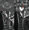
Figure 1. Axial CT of the cervical spine (A) in a 30-year-old trauma patient status post MVA demonstrating C1 anterior and posterior arch fractures (red arrows) and right lateral mass fracture. Axial T2-weighted MRI (B) in the same patient demonstrating a left transverse atlantal ligament tear (red arrowheads). Sagittal T2-weighted (C) and STIR-weighted (D) MRI of the cervical spine in the same patient demonstrating low-grade anterior and posterior atlanto-occipital membrane complex injuries (red arrows) with otherwise intact major craniocervical junction ligaments. Anterior and posterior atlanto-occipital membrane complex tears were relatively common in the setting of transverse atlantal ligament tears.
CT: computed tomography; MVA: motor vehicle accident; MRI: magnetic resonance imaging; STIR: short tau inversion recovery
Figure 2
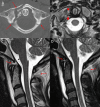
Figure 2. Axial CT of the cervical spine (A) in a 32-year-old trauma patient status post MVA demonstrating comminuted, left C1 anterior and posterior arch fractures (red arrows). Axial T2-weighted MRI (B) in the same patient demonstrating a left transverse atlantal ligament tear (red arrowheads). Sagittal T2-weighted (C) and STIR-weighted (D) MRI of the cervical spine in the same patient demonstrating low-grade anterior and posterior atlanto-occipital membrane complex injuries (red arrows) with otherwise intact major craniocervical junction ligaments. Anterior and posterior atlanto-occipital membrane complex tears were relatively common in the setting of transverse atlantal ligament tears.
CT: computed tomography; MVA: motor vehicle accident; MRI: magnetic resonance imaging; STIR: short tau inversion recovery
Figure 3
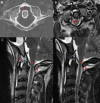
Figure 3. Axial CT of the cervical spine (A) in an 18-year-old trauma patient status post MVA demonstrating comminuted, right C1 anterior arch fracture and bilateral posterior arch fractures (red arrows). Axial T2-weighted MRI (B) in the same patient demonstrating a right transverse atlantal ligament tear (red arrowheads). Sagittal T2-weighted (C) and STIR-weighted (D) MRI of the cervical spine in the same patient demonstrating anterior and posterior atlanto-occipital membrane complex injuries (red arrows) with otherwise intact major craniocervical junction ligaments. Anterior and posterior atlanto-occipital membrane complex tears were relatively common in the setting of transverse atlantal ligament tears.
CT: computed tomography; MVA: motor vehicle accident; MRI: magnetic resonance imaging; STIR: short tau inversion recovery
Figure 4
Figure 4. Axial CT of the cervical spine (A) in a 48-year-old trauma patient status post MVA demonstrating widening of the anterior atlanto-dens interval (red line) without a C1 fracture. Axial T2-weighted MRI (B) in the same patient demonstrating a central transverse atlantal ligament tear (red arrow). Sagittal T2-weighted (C) and STIR-weighted (D) MRI of the cervical spine in the same patient demonstrating subclival tectorial membrane tear (red arrows), right alar ligament tear (blue arrowheads), and anterior and posterior atlanto-occipital membrane complex tears (red stars). The majority of observed patients in our study with atlantoaxial instability did not have major craniocervical junction ligament tears.
CT: computed tomography; MVA: motor vehicle accident; MRI: magnetic resonance imaging; STIR: short tau inversion recovery

References

    1. The spectrum of traumatic injuries at the craniocervical junction: a review of imaging findings and management. Siddiqui J, Grover PJ, Makalanda HL, Campion T, Bull J, Adams A. Emerg Radiol. 2017;24:377–385. - PubMed
    1. Radiologic spectrum of craniocervical distraction injuries. Deliganis AV, Baxter AB, Hanson JA, Fisher DJ, Cohen WA, Wilson AJ, Mann FA. Radiographics. 2000;20:0–50. - PubMed
    1. The anterior atlantodental ligament: its anatomy and potential functional significance. Tubbs RS, Mortazavi MM, Louis RG, et al. World Neurosurg. 2012;77:775–777. - PubMed
    1. Anatomic, functional, and radiographic review of the ligaments of the craniocervical junction. Fiester P, Rao D, Soule E, Orallo P, Rahmathulla G. J Craniovertebr Junction Spine. 2021;12:4–9. - PMC - PubMed
    1. Ligaments of the craniocervical junction. Tubbs RS, Hallock JD, Radcliff V, et al. J Neurosurg Spine. 2011;14:697–709. - PubMed

LinkOut - more resources