Review
. 2022 Dec 21;289(1989):20222054.
doi: 10.1098/rspb.2022.2054. Epub 2022 Dec 14.

Drosophila olfaction: past, present and future

Affiliations
Review

Drosophila olfaction: past, present and future

Richard Benton. Proc Biol Sci. .

Abstract

Among the many wonders of nature, the sense of smell of the fly Drosophila melanogaster might seem, at first glance, of esoteric interest. Nevertheless, for over a century, the 'nose' of this insect has been an extraordinary system to explore questions in animal behaviour, ecology and evolution, neuroscience, physiology and molecular genetics. The insights gained are relevant for our understanding of the sensory biology of vertebrates, including humans, and other insect species, encompassing those detrimental to human health. Here, I present an overview of our current knowledge of D. melanogaster olfaction, from molecules to behaviours, with an emphasis on the historical motivations of studies and illustration of how technical innovations have enabled advances. I also highlight some of the pressing and long-term questions.

Keywords: Drosophila melanogaster; animal behaviour; neural circuit; neurophysiology; olfaction; olfactory receptor.


Conflict of interest statement

I declare I have no competing interests.

Figures

Figure 1.
Figure 1.
Olfactory organs. (a) Scanning electron micrograph (SEM) of the head of adult D. melanogaster, showing the two bilaterally symmetric olfactory organs. Adapted from [22] (copyright © Cold Spring Harbor Laboratory Press). (b) SEM of a D. melanogaster antenna, illustrating the dense array of morphologically diverse sensilla (which house olfactory sensory neuron dendrites) covering the surface. Scale bar, 50 µm. Adapted from [23].
Figure 2.
Figure 2.
Olfactory receptors. Model of a hypothetical heterotetrameric complex of D. melanogaster Or22a and the co-receptor Orco (two subunits each). The approximate position of the plasma membrane is indicated in the side view. In Or22a subunits, the residue highlighted in white (M93) is a major contributor to defining behaviourally relevant odour response differences between D. melanogaster and D. sechellia Or22a orthologues [34]; this residue is located within the putative odour-binding site [35]. The ion channel pore is formed at the interface of all four subunits [36]. Models of protein monomers were predicted by AlphaFold2 [37,38]; these exhibit very strong similarity to cryo-electron microscopic (cryoEM) structures of Ors from other insects [35,36]. Models were aligned to the cryoEM structure of the Orco homotetramer from the fig wasp (Apocrypta bakeri) [36] using Coot [39] and visualized in PyMol v. 2.5.4. Although the stoichiometry of Or/Orco complexes is unknown, evidence suggests that they contain at least two tuning Or subunits [32,40].
Figure 3.
Figure 3.
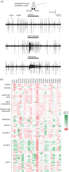
Olfactory function. (a) Electrophysiological recordings from the antennal basiconic sensillum 3 (ab3), which houses two neurons (ab3A and ab3B) that express Or22a/b and Or85b, respectively. The neurons can be distinguished both by ‘spike’ (action potential) amplitude and their responses to different odours (diluted to 0.001% v/v in the paraffin oil solvent and presented during 1 s). Adapted from [22] (copyright © Cold Spring Harbor Laboratory Press). (b) Combinatorial coding of odours by Ors: the first large-scale profiling of responses of many Ors to a chemically diverse panel of stimuli. Here, Ors were transgenically expressed in the ‘empty’ Or22a/b neuron (lacking the endogenous receptors) to provide a consistent cellular background for comparison of receptor function. Data are replotted from [61]; the scale is shown on the right. Negative responses reflect odours that decrease the basal spiking frequency of neurons. Some receptors for which no strong agonists were identified (e.g. Or47b) were later found to respond to pheromones [67,68].
Figure 4.
Figure 4.
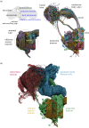
Olfactory circuits. (a) Top left: schematic of the principal paths of olfactory information flow in the D. melanogaster brain (outlined in grey), indicating the main neuron classes and brain regions. The circuitry is bilaterally symmetric (and most olfactory sensory neurons (OSNs) project to both antennal lobes) but only one hemibrain pathway is illustrated. Below are electron microscopic-resolution connectomic reconstructions of OSNs (including some antennal hygrosensory and thermosensory neurons [–86]; contralateral innervations are cut off on the right side) and uniglomerular projection neurons (PNs) in the antennal lobe. Partner OSNs and PNs, converging on a common glomerulus, are colour-matched; one such glomerulus is highlighted in both images. For this PN class, a subset of the soma (located outside the lobe) are indicated with white arrowheads; black arrowheads point to the axons that project to the higher brain centres. (b) Connectomic reconstructions of the indicated neuronal populations. Data in (a,b) are adapted from [87], prepared by P. Schlegel; note that missing surface ‘strips’ in the middle of the antennal lobe are due to absent data.
Figure 5.
Figure 5.
Olfactory behaviour. (a) Historical tracking: manually traced paths of individual walking D. ampelophila (the former name for D. melanogaster) from the edge of a 5 × 5 inch arena toward a piece of fermenting banana at the centre. Reproduced from [1], with permission. (b) State-of-the-art tracking: schematic of a wind tunnel through which a controlled odour plume can be introduced; a multi-camera system enables automated three-dimensional tracking of flies interacting with this plume in flight. On the right is the trajectory (top and side views) of D. melanogaster in a continuous plume of ethanol. Grey dashed lines on the left indicate the upwind wall of the tunnel towards which the animal flies. The trajectories are colour-coded for the instantaneous concentration of odour at a given point in the plume (as measured using a photoionization detector); the reconstructed odour experience of the animal over the course of its flight is plotted below the trajectories. Two time-synchronized reference points are indicated with orange and green arrowheads. Adapted from [214], with permission.

References

    1. Barrows WM. 1907. The reactions of the pomace fly, Drosophila ampelophila Loew, to odorous substances. J. Exp. Zool. 4, 515-537. (10.1002/jez.1400040403) - DOI
    1. Greenspan RJ, Ferveur JF. 2000. Courtship in Drosophila. Annu. Rev. Genet. 34, 205-232. (10.1146/annurev.genet.34.1.205) - DOI - PubMed
    1. Quinn WG, Greenspan RJ. 1984. Learning and courtship in Drosophila: two stories with mutants. Annu. Rev. Neurosci. 7, 67-93. (10.1146/annurev.ne.07.030184.000435) - DOI - PubMed
    1. Weiner J. 1999. Time, love, memory: a great biologist and his quest for the origins of behavior. New York, NY: Knopf.
    1. Stocker RF. 1994. The organization of the chemosensory system in Drosophila melanogaster: a review. Cell Tissue Res. 275, 3-26. (10.1007/BF00305372) - DOI - PubMed

Publication types

LinkOut - more resources