. 2023 Apr;102(4):412-421.
doi: 10.1177/00220345221138533. Epub 2022 Dec 14.

Noninvasive Monitoring of Radiation-Induced Salivary Gland Vascular Injury

Affiliations

Noninvasive Monitoring of Radiation-Induced Salivary Gland Vascular Injury

E R Bolookat et al. J Dent Res. 2023 Apr.

Abstract

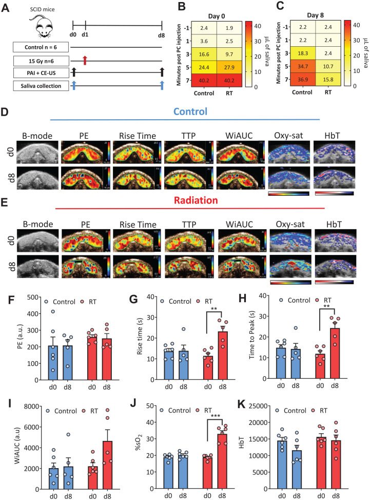
Xerostomia is a common side effect of radiation therapy (RT) in patients with head and neck cancer. However, limited information is available on the temporal dynamics of parenchymal and vascular changes in salivary glands following RT. To address this gap in knowledge, we conducted experimental studies in mice employing ultrasound (US) with coregistered photoacoustic imaging (PAI) to noninvasively assess the early and late changes in salivary gland size, structure, vascularity, and oxygenation dynamics following RT. Multiparametric US-PAI of salivary glands was performed in immune-deficient and immune-competent mice before and after RT along with correlative sialometry and ex vivo histologic-immunohistochemical validation. US revealed reduction in gland volume and an early increase in vascular resistance postradiation. This was accompanied by a reduction in glandular oxygen consumption on PAI. Imaging data correlated strongly with salivary secretion and histologic evidence of acinar damage. The magnitude and kinetics of radiation response were impacted by host immune status, with immunodeficient mice showing early and more pronounced vascular injury and DNA damage response compared to immunocompetent animals. Our findings demonstrate the ability of noninvasive US-PAI to monitor dynamic changes in salivary gland hemodynamics following radiation and highlight the impact of the host immune status on salivary gland radiation injury.

Keywords: molecular imaging; radiation; saliva; ultrasonography; xerostomia.


Conflict of interest statement

The authors declared the following potential conflicts of interest with respect to the research, authorship, and/or publication of this article: L.J. Rich is currently an employee of Fujifilm-VisualSonics Corporation. The remaining authors declare no competing financial interest. The funding sponsors had no role in the design of the study, collection, analyses, or interpretation of data; writing of the manuscript; and the decision to publish the results.

Figures

Figure 1.
Figure 1.
Sonographic and photoacoustic imaging of early salivary gland response to radiation (RT). (A) Study design: naive severe combined immunodeficient (SCID) mice were separated into control (n = 4) and fractionated radiation (fRT; 6 Gy × 5; n = 6) or single-dose (RT; 30 Gy; n = 6) arms. Animals underwent ultrasound with coregistered photoacoustic imaging (US-PAI) before (d0) and 1 wk after radiation treatment (d12) along with correlative sialometry. Salivary glands were collected for histologic evaluation following imaging. (B) B-mode US images (top) and corresponding 3-dimensional volume renderings (bottom) of salivary glands from a representative animal in the control, fRT, and RT arms on d0 and d12. (C) Change in US-based salivary gland volumes in control and radiated animals on d0 and d12. (D) Wet gland weights of salivary glands from animals in control, fRT, and RT groups on d12. (E) Correlation plot between US-based gland volumes and wet gland weights between control and irradiated animals on d12. (F) Heatmap of secreted saliva volume (µL) in response to pilocarpine (PC; 20 mg/kg, intraperitoneally; y-axis represents minutes after pilocarpine) stimulation showing significant reduction in saliva secretion on d12 in irradiated animals (P < 0.0001 at 5 and 7 min post-PC) compared to controls. Mean saliva volume at 1, 3, 5, and 7 min after pilocarpine injection at each study time point (x-axis) is shown. Parametric maps of hemoglobin concentration (HbT; G) and percent oxygen saturation (%sO2; H) overlaid on the B-mode US images of salivary glands are shown for a representative animal from the control and radiation arms at baseline (day 0) and 1 wk posttreatment (day 12). Corresponding bar graphs of quantitative estimates for HbT (I) and %sO2 (J) for animals in the 3 groups are shown. Panel of photomicrographs represents hematoxylin and eosin (H&E; K) and Masson’s trichrome (L) stained sections of control and radiated glands on d12. Images are shown at 20× magnification (scale bar 100 µm); inset: whole gland images. Control glands showed intact architecture with serous acini containing secretory granules (H&E; green arrow) and minimal collagen (blue; Masson’s trichrome). Glands from animals in both fRT and RT arms revealed shrinkage of acini (black arrows) with hyperchromatic nuclei (yellow arrows) and shrinkage of ducts with coarse eosinophilic granules. Quantitative estimates of percent acinar coverage (M) and acinar size (N) from H&E-stained sections and collagen density based on Masson’s trichrome staining (O) for animals in all 3 groups. *P < 0.05. **P < 0.01. ***P < 0.001. ****P < 0.0001.
Figure 2.
Figure 2.
Combined contrast-enhanced ultrasound (CE-US) and photoacoustic imaging (PAI) of radiation-induced salivary gland vascular injury. (A) Study design: naive severe combined immunodeficient (SCID) mice were separated into control (n = 6) and radiation (15 Gy; n = 6) arms. Animals underwent combined contrast-enhanced US and PAI examination before (d0) and 1 wk after radiation treatment (d8) along with measurement of stimulated saliva secretion after pilocarpine administration (10 mg/kg, intraperitoneally). Heatmaps of saliva secretion (µL) before and after pilocarpine administration in control (B) and radiation (C) cohorts at baseline (d0) and 1 wk after radiation (d8). Spatially coregistered parametric maps of peak enhancement (PE), rise time, time to peak (TTP), wash-in area under the curve (WiAUC), oxygen saturation (Oxy-sat), and hemoglobin concentration (HbT) on d0 and d8 for control (D) and radiated glands (E). The corresponding B-mode image of the glands is also shown on the left. Bar graphs showing CE-US–based estimates of PE (F), rise time (G), TTP (H), WiAUC (I), and PAI-based estimates of percent oxygen saturation (%sO2) (J) and HbT (K). **P < 0.01. ***P < 0.001.
Figure 3.
Figure 3.
Sonographic assessment of long-term radiation (RT) injury to salivary glands in immunodeficient and immunocompetent mice. (A) Study design: female severe combined immunodeficient (SCID) mice (SCID, n = 6; RT, n = 10) and immunocompetent Bl6 mice (control, n = 4; RT, n = 7) were separated into control and radiation (17.5 Gy) arms. Longitudinal ultrasound–photoacoustic imaging (US-PAI) with correlative sialometry (pilocarpine [PC], 10 mg/kg, intraperitoneally) was performed at baseline, 1 wk (day 7), 4 wk (day 28), and 8 wk (day 56) after radiation. (B) US images of salivary glands in control and radiated animals of both strains at baseline and days 7, 28, and 56. Change in gland volumes calculated from 3-dimensional US over the study period for SCID (C) and albino Bl6 (D). Heatmaps of saliva secretion (µL) before and after PC administration in SCID (E, F) and albino Bl6 (G, H) mice at baseline and days 7, 28, and 56 after radiation. *P < 0.05. **P < 0.01. ***P < 0.001.
Figure 4.
Figure 4.
Photoacoustic imaging of radiation-induced vascular injury to salivary glands in immunodeficient and immunocompetent hosts. Parametric maps of oxygen saturation (Oxy-Sat) and hemoglobin concentration (HbT) of control and radiated glands (17.5 Gy) in immunodeficient severe combined immunodeficient (SCID) (A) and immunocompetent Bl6 (D) mice at baseline, 1 wk (day 7), 4 wk (day 28), and 8 wk (day 56) after radiation. Pseudo-colorized images overlaid on the B-mode ultrasound (US) of the salivary glands are shown. Box-and-whisker plots showing change in percent oxygen saturation (%sO2) (B, E) and HbT (C, F) levels in salivary glands for control and radiated animals in both strains over the 8-wk study period. *P < 0.05. **P < 0.01. ***P < 0.001.
Figure 5.
Figure 5.
Histologic and immunohistochemical validation of radiation-induced salivary gland vascular injury and DNA damage in immunodeficient and immunocompetent hosts. Photomicrographs of hematoxylin and eosin (H&E), CD31, and phosphorylated γH2AX immunostained sections (20× magnification; scale bar 100 µm) of salivary glands from control and radiated severe combined immunodeficient (SCID) (A) and immunocompetent Bl6 (E) mice at 4 wk (day 28) and 8 wk (day 56) after radiation (RT). Glandular atrophy (green arrows) was seen on d28 and d56 after RT along with vacuolization (orange arrows) and fibrosis (white arrows) compared to control animals in both strains. H&E of Bl6 mice showed large nuclei and acinar atrophy (green arrow) on d28 and vacuolization (yellow arrows) on d56. A significant increase (P < 0.01) in phospho-γH2AX staining (black arrows) was seen after RT in SCID mice. Bar graphs showing CD31+-based vessel counts (B, F) and lumen size (C, G) and DNA damage (γH2AX score; D, H) of salivary glands for control and radiated animals in both strains. *P < 0.05. **P < 0.01. ***P < 0.001.

References

    1. Biedermann KA, Sun JR, Giaccia AJ, Tosto LM, Brown JM. 1991. Scid mutation in mice confers hypersensitivity to ionizing radiation and a deficiency in DNA double-strand break repair. Proc Natl Acad Sci U S A. 88(4):1394–1397. - PMC - PubMed
    1. Burdelya LG, Gleiberman AS, Toshkov I, Aygun-Sunar S, Bapardekar M, Manderscheid-Kern P, Bellnier D, Krivokrysenko VI, Feinstein E, Gudkov AV. 2012. Toll-like receptor 5 agonist protects mice from dermatitis and oral mucositis caused by local radiation: implications for head-and-neck cancer radiotherapy. Int J Radiat Oncol Biol Phys. 83(1):228–234. - PMC - PubMed
    1. Chambers MS, Garden AS, Kies MS, Martin JW. 2004. Radiation-induced xerostomia in patients with head and neck cancer: pathogenesis, impact on quality of life, and management. Head Neck. 26(9):796–807. - PubMed
    1. Choi JH, Wu HG, Jung KC, Lee SH, Kwon EK. 2009. Apoptosis and expression of AQP5 and TGF-beta in the irradiated rat submandibular gland. Cancer Res Treat. 41(3):145–154. - PMC - PubMed
    1. Cotrim AP, Sowers A, Mitchell JB, Baum BJ. 2007. Prevention of irradiation-induced salivary hypofunction by microvessel protection in mouse salivary glands. Mol Ther. 15(12):2101–2106. - PubMed

Publication types