. 2023 Mar;29(3):352-362.
doi: 10.1177/13524585221141964. Epub 2022 Dec 14.

Relationship between paramagnetic rim lesions and slowly expanding lesions in multiple sclerosis

Affiliations

Relationship between paramagnetic rim lesions and slowly expanding lesions in multiple sclerosis

Alberto Calvi et al. Mult Scler. 2023 Mar.

Abstract

Background: Magnetic resonance imaging (MRI) markers for chronic active lesions in MS include slowly expanding lesions (SELs) and paramagnetic rim lesions (PRLs).

Objectives: To identify the relationship between SELs and PRLs in MS, and their association with disability.

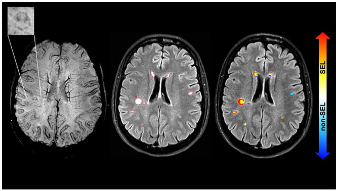
Methods: 61 people with MS (pwMS) followed retrospectively with MRI including baseline susceptibility-weighted imaging, and longitudinal T1 and T2-weighted scans. SELs were computed using deformation field maps; PRLs were visually identified. Mixed-effects models assessed differences in Expanded Disability Status Scale (EDSS) score changes between the group defined by the presence of SELs and or PRLs.

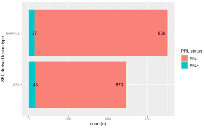
Results: The median follow-up time was 3.2 years. At baseline, out of 1492 lesions, 616 were classified as SELs, and 80 as PRLs. 92% of patients had ⩾ 1 SEL, 56% had ⩾ 1 PRL, while both were found in 51%. SELs compared to non-SELs were more likely to also be PRLs (7% vs. 4%, p = 0.027). PRL counts positively correlated with SEL counts (ρ= 0.28, p = 0.03). SEL + PRL + patients had greater increases in EDSS over time (beta = 0.15/year, 95% confidence interval (0.04, 0.27), p = 0.009) than SEL+PRL-patients.

Conclusion: SELs are more numerous than PRLs in pwMS. Compared with either SELs or PRLs found in isolation, their joint occurrence was associated with greater clinical progression.

Keywords: Chronic active lesions; multiple sclerosis; paramagnetic rim lesions (PRLs); slowly expanding lesions (SELs); susceptibility-weighted imaging (SWI); volumetric MRI.


Conflict of interest statement

The author(s) declared no potential conflicts of interest with respect to the research, authorship, and/or publication of this article.

Figures

Figure 1.
Figure 1.
Stacked bar plot showing the total number of lesions classified as SEL or non-SEL, and the correspondent number of PRL within each subtype. The figure, drawn with R, shows the counts of lesions divided between the SEL and the non-SEL categories, and the corresponding PRLs in each subtype. The proportion of PRLs was higher among the SELs than among the non-SELs (7% vs. 4%, p = 0.027).
Figure 2.
Figure 2.
Example of a PRL at baseline and the correspondence to a SEL. The images show from left to right: SWI at baseline (the top left rectangle shows a magnification of the paramagnetic rim lesion), FLAIR with lesion mask countered in red, Jacobian map superimposed indicating that the PRL corresponds to a SEL. The images are from a 45-year-old person with a CIS and the MRI was performed 4 months after onset. At baseline the EDSS was 0 and last follow-up (~4 years) was 1.5. CIS: clinically isolated syndrome; EDSS: Expanded Disability Status Scale; PRL: paramagnetic rim lesion; FLAIR: fluid-attenuated inversion recovery; PRL: paramagnetic rim lesion; SWI: susceptibility-weighted imaging; SEL: slowly expanding lesion.

References

    1. Luchetti S, Fransen NL, van Eden CG, et al.. Progressive multiple sclerosis patients show substantial lesion activity that correlates with clinical disease severity and sex: A retrospective autopsy cohort analysis. Acta Neuropathol 2018; 135(4): 511–528. - PMC - PubMed
    1. Frischer JM, Weigand SD, Guo Y, et al.. Clinical and pathological insights into the dynamic nature of the white matter multiple sclerosis plaque. Ann Neurol 2015; 78(5): 710–721. - PMC - PubMed
    1. Bagnato F, Hametner S, Yao B, et al.. Tracking iron in multiple sclerosis: A combined imaging and histopathological study at 7 tesla. Brain 2011; 134(Pt. 12): 3602–3615. - PMC - PubMed
    1. Popescu BF, Frischer JM, Webb SM, et al.. Pathogenic implications of distinct patterns of iron and zinc in chronic MS lesions. Acta Neuropathol 2017; 134(1): 45–64. - PMC - PubMed
    1. Yao B, Ikonomidou VN, Cantor FK, et al.. Heterogeneity of multiple sclerosis white matter lesions detected with T2*-weighted imaging at 7.0 tesla. J Neuroimaging 2015; 25(5): 799–806. - PMC - PubMed