. 2023 Jan;27(1):113-126.
doi: 10.1111/jcmm.17643. Epub 2022 Dec 14.

Circ_0000052/miR-382-3p axis induces PD-L1 expression and regulates cell proliferation and immune evasion in head and neck squamous cell carcinoma

Affiliations

Circ_0000052/miR-382-3p axis induces PD-L1 expression and regulates cell proliferation and immune evasion in head and neck squamous cell carcinoma

De-Jun Zhang et al. J Cell Mol Med. 2023 Jan.

Abstract

A better understanding of the mechanisms underlying PD-L1 aberrant expression in head and neck squamous cell carcinoma (HNSCC) will help reveal predictive biomarkers and overcome resistance to treatment. In this study, the prognostic significance of PD-L1 in forty-five HNSCC archival samples was determined by qRT-PCR. The biological function associated with malignant behaviour was assessed by PD-L1 depletion, miR-382-3p re-expression and regulation of circ_0000052. The interactions of PD-L1-miRNA and miRNA-circRNA were determined by qRT-PCR, Western blot analysis, dual-luciferase reporter assays and RNA immunoprecipitation assays. PD-L1 was highly expressed in patient samples and cancer cell lines. Higher levels of PD-L1 were associated with patient recurrences and play a pivotal role in regulating cell proliferation, migration, invasion, clonogenicity and apoptosis. In addition to demonstrating that the IFN-γ/JAK2/STAT1 signalling pathway can induce PD-L1 overexpression in HNSCC, a novel mechanism by which upregulated circ_0000052 mediates PD-L1 overexpression was also demonstrated. To do this, circ_0000052 competitively binds to miR-382-3p and alleviates its repression of PD-L1. This leads to overexpression of PD-L1, causing the aggressiveness of the cells. Our data demonstrate that circ_0000052 is oncogenic, and the circ_0000052/miR-382-3p/PD-L1 axis is critical in HNSCC progression. The manipulation of circRNAs/miRNAs in combination with anti-PD-L1 therapy may improve personalized disease management.

Keywords: PD-L1; circular RNA; head and neck squamous cell carcinoma; immunotherapy; microRNA.


Conflict of interest statement

The authors declare no conflict of interest.

Figures

FIGURE 1
FIGURE 1
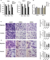
PD‐L1 overexpression is associated with poor prognosis in HNSCC patients. (A) The expression of PD‐L1 was increased in HNSCC examined by RT‐qPCR. (B) Higher PD‐L1 expression was associated with worse overall survival as analysed by Kaplan–Meier survival analysis and the log‐rank test (p < 0.05). (C) PD‐L1 overexpression in two HNSCC cell lines. (D) siRNAs effectively deplete PD‐L1. * p < 0.05; ** p < 0.01.
FIGURE 2
FIGURE 2
PD‐L1 depletion reduces cell proliferation, migration and invasion while increasing apoptosis activity. (A) siRNA for PD‐L1 was transfected into Fadu and SCC‐9 cells, respectively, and cell viability was determined using the MTS assay after 24–72 h post‐transfection. (B) Caspase‐3/7 activities were assessed for Fadu and SCC‐9 cells. The cells were transfected with siRNA1 (40 nM) for PD‐L1 or control siRNA, and the activities of caspases were measured 24 and 48 h post‐transfection. The data are plotted as the average relative activity. (C and D) Representative images and quantification of the reduction in migratory capacity (top panel) and invasion (bottom panel) of Fadu and SCC‐9 cells that were transfected with 40 nM of siRNA1 or siRNA2 for PD‐L1, compared to negative scramble controls. All the data were generated from three independent experiments. *p < 0.05; **p < 0.01.
FIGURE 3
FIGURE 3
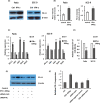
Interferon‐γ (IFN‐γ) induces PD‐L1 expression in HNSCC. (A and B) PD‐L1 expression levels were measured in Fadu and SCC‐9 cell lines 72 h after treatment with or without IFN‐ γ (100 ng). (C) JAK/STAT signalling pathway gene expression levels in cell lines 48 h after control or 100 ng IFN‐γ treatment. The levels of gene expression are normalized to β‐actin and GAPDH. (D) Expression of miR‐382‐3p in cell lines following IFN‐γ treatment. (E and F) Expression of PD‐L1 in Fadu cells was stimulated by IFN‐γ and Stat1. Each experiment was repeated at least three times. *p < 0.05, **p < 0.01.
FIGURE 4
FIGURE 4
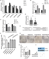
miR‐382‐3p directly targets PD‐L1 in HNSCC. (A) qRT‐PCR validation of five putative miRNA targets for PD‐L1 identified through bioinformatics analysis. (B) Mimics of miRNAs increased the expression of miR‐382‐3p and miR‐375‐5p in Fadu and SCC‐9 cells at 48 h post‐transfection. (C) PD‐L1 expression was reduced by transfection of premiR‐382‐3p and premiR‐375‐5p. (D) MiR‐382‐3p expression levels in HNSCC tissues were measured. (E) The targeting sites of the wide type (WT) or mutation (mut) of PD‐L1 with miR‐382‐3p. (F) Luciferase reporter assays in Fadu and SCC‐9 cells co‐transfected with pMIR‐PD‐L1 or pMIR‐PD‐L1‐mut plasmids, scramble negative control miR, and either premiR‐382‐3p or premiR‐375‐5p. Samples were analysed 72 h post‐transfection, and data were normalized to the pMIR report only transfection. (G and H) Colony formation assay of Fadu cells transfected with siRNA‐PD‐L1, premiR‐382‐3p or antimiR‐382‐3p. (I) PD‐L1 expression levels were regulated by miR‐382‐3p, as detected by Western blotting. Data represent the mean ± SE, n = 3. *p < 0.05, **p < 0.01.
FIGURE 5
FIGURE 5
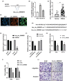
Circ_0000052 regulates cell growth and invasion by targeting miR‐382‐3p. (A) A schematic representation of circ_0000052 and its optimally transcribed gene. (B and C) The expression level of circ_0000052 was detected in HNSCC cells and tissue specimens. (D) The cellular distribution of circ_0000052 was analysed by fluorescence in situ hybridization (FISH). Nuclei were stained with DAPI (blue). Orange indicates circ_0000052. Scale bar = 50 μm. (E) The stability of circ_0000052 was determined by qRT‐PCR after treatment with RNase R or mock in total RNAs derived from Fadu cells. (F) Luciferase activity in Fadu cells co‐transfected with luciferase reporters containing miR‐382‐3p sequences with wild‐type or mutated circ_0000052 binding sites and the mimics of miR‐382‐3p or control. (G) Schema of the predicted binding site of miR‐382‐3p on the wide‐type (WT) of circ_0000052. A mutant sequence of circ_0000052 was shown. (H‐J) Depletion of circ_0000052 reduced cell viability, migratory capacity and cell invasion. All data were represented as means ± SE, n = 3. *p < 0.05; **p < 0.01.
FIGURE 6
FIGURE 6
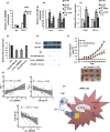
Circ_0000052 interacts with PD‐L1 via sponge miR‐382‐3p. (A) The RIP assay was executed in Fadu cells after introducing enriched miR‐382‐3p, followed by detecting the expression of circ_0000052. (B and C) After co‐transfection of cells with premiR‐382‐3p or overexpressed circ_0000052 plasmids or both, the expression levels of miR‐382‐3p or PD‐L1 were determined. (D and E) Rescue experiments were performed by transfecting premiR‐382‐3p or si‐circ_0000052 or co‐transfecting both. PD‐L1 levels were detected by qRT‐PCR and Western blotting. (F) Enhanced miR‐382‐3p and depletion of circ_0000052 delayed tumour growth compared with the control. n = 3/group. Tumour volume was measured. (G) The correlation between PD‐L1 and circ_0000052, PD‐L1 and miR‐382‐3p, circ_0000052 and miR‐382‐3p was analysed by Pearson correlation analysis. *p < 0.05, **p < 0.01. (H) A schematic diagram to illustrate a hypothetical model in which circ_0000052 acts as a competitive ceRNA to sponge‐adsorb miR‐382‐3p and alleviate the inhibitory effect on PD‐L1 through a circRNA‐sponge mechanism.

References

    1. Argiris A, Karamouzis MV, Raben D, Ferris RL. Head and neck cancer. Lancet. 2008;371:1695‐1709. - PMC - PubMed
    1. Leemans CR, Braakhuis BJ, Brakenhoff RH. The molecular biology of head and neck cancer. Nat Rev Cancer. 2011;11:9‐22. - PubMed
    1. Shibata H, Saito S, Uppaluri R. Immunotherapy for head and neck cancer: A paradigm shift from induction chemotherapy to Neoadjuvant immunotherapy. Front Oncol. 2021;11:727433. - PMC - PubMed
    1. Prestipino A, Zeiser R. Clinical implications of tumor‐intrinsic mechanisms regulating PD‐L1. Sci Transl Med. 2019;11:eaav4810. - PubMed
    1. Cui P, Jing P, Liu X, Xu W. Prognostic significance of PD‐L1 expression and its tumor‐intrinsic functions in Hypopharyngeal squamous cell carcinoma. Cancer Manag Res. 2020;12:5893‐5902. - PMC - PubMed

Publication types