

Abnormal echocardiographic findings after COVID-19 infection: a multicenter registry
- PMID: 36515755
- PMCID: PMC9376039
- DOI: 10.1007/s10554-022-02706-9
Abnormal echocardiographic findings after COVID-19 infection: a multicenter registry
Abstract
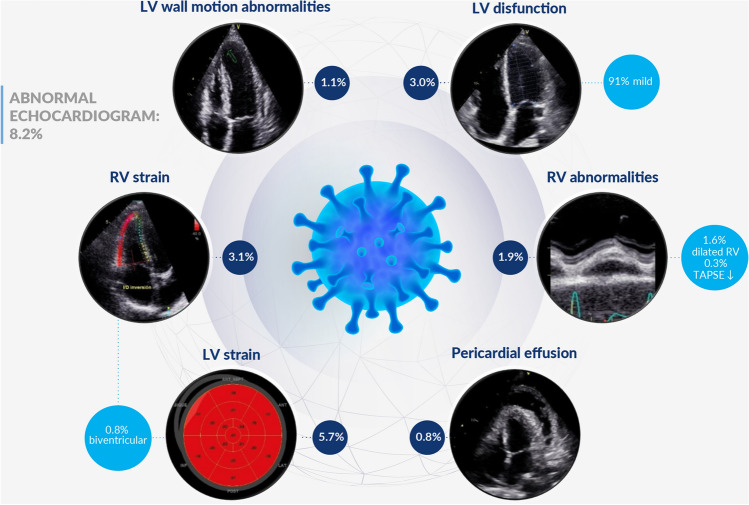
The Coronavirus Disease 2019 (COVID-19) pandemic has transformed health systems worldwide. There is conflicting data regarding the degree of cardiovascular involvement following infection. A registry was designed to evaluate the prevalence of echocardiographic abnormalities in adults recovered from COVID-19. We prospectively evaluated 595 participants (mean age 45.5 ± 14.9 years; 50.8% female) from 10 institutions in Argentina and Brazil. Median time between infection and evaluation was two months, and 82.5% of participants were not hospitalized for their infection. Echocardiographic studies were conducted with General Electric equipment; 2DE imaging and global longitudinal strain (GLS) of both ventricles were performed. A total of 61.7% of the participants denied relevant cardiovascular history and 41.8% had prolonged symptoms after resolution of COVID-19 infection. Mean left ventricular ejection fraction (LVEF) was 61.0 ± 5.5% overall. In patients without prior comorbidities, 8.2% had some echocardiographic abnormality: 5.7% had reduced GLS, 3.0% had a LVEF below normal range, and 1.1% had wall motion abnormalities. The right ventricle (RV) was dilated in 1.6% of participants, 3.1% had a reduced GLS, and 0.27% had reduced RV function. Mild pericardial effusion was observed in 0.82% of participants. Male patients were more likely to have new echocardiographic abnormalities (OR 2.82, p = 0.002). Time elapsed since infection resolution (p = 0.245), presence of symptoms (p = 0.927), or history of hospitalization during infection (p = 0.671) did not have any correlation with echocardiographic abnormalities. Cardiovascular abnormalities after COVID-19 infection are rare and usually mild, especially following mild infection, being a low GLS of left and right ventricle, the most common ones in our registry. Post COVID cardiac abnormalities may be more frequent among males.
Keywords: COVID-19; Diagnostic imaging; Echocardiography; Humans; Myocarditis; Ventricular remodeling.
© 2022. The Author(s), under exclusive licence to Springer Nature B.V.
Conflict of interest statement
The authors declare no competing interests.
Figures
References
Publication types
MeSH terms
LinkOut - more resources
Full Text Sources
Medical
Miscellaneous
