. 2023 Jan 12;66(1):804-821.
doi: 10.1021/acs.jmedchem.2c01693. Epub 2022 Dec 14.

The Identification of Potent, Selective, and Brain Penetrant PI5P4Kγ Inhibitors as In Vivo-Ready Tool Molecules

Affiliations

The Identification of Potent, Selective, and Brain Penetrant PI5P4Kγ Inhibitors as In Vivo-Ready Tool Molecules

Timothy P C Rooney et al. J Med Chem. .

Abstract

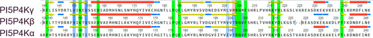
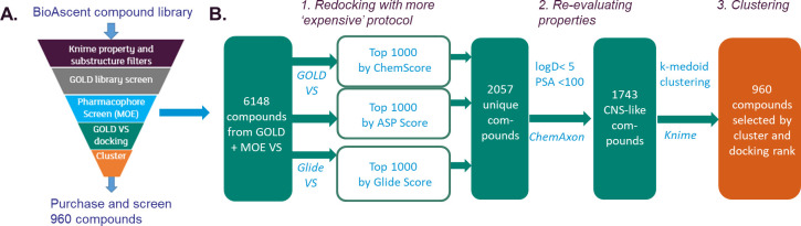
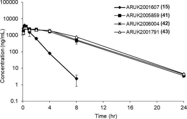
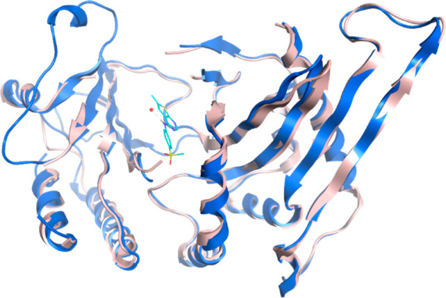
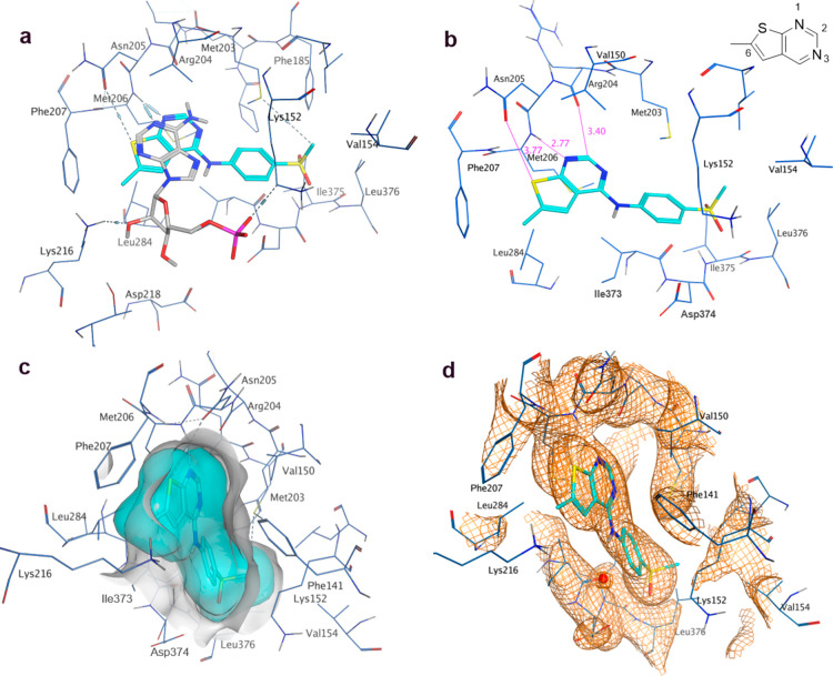
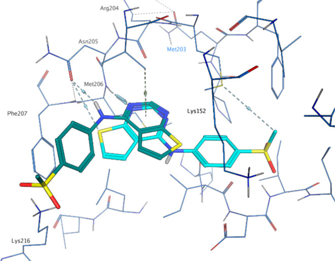
Owing to their central role in regulating cell signaling pathways, the phosphatidylinositol 5-phosphate 4-kinases (PI5P4Ks) are attractive therapeutic targets in diseases such as cancer, neurodegeneration, and immunological disorders. Until now, tool molecules for these kinases have been either limited in potency or isoform selectivity, which has hampered further investigation of biology and drug development. Herein we describe the virtual screening workflow which identified a series of thienylpyrimidines as PI5P4Kγ-selective inhibitors, as well as the medicinal chemistry optimization of this chemotype, to provide potent and selective tool molecules for further use. In vivo pharmacokinetics data are presented for exemplar tool molecules, along with an X-ray structure for ARUK2001607 (15) in complex with PI5P4Kγ, along with its selectivity data against >150 kinases and a Cerep safety panel.


Conflict of interest statement

The authors declare no competing financial interest.

Figures

Figure 1
Figure 1
Sequence alignment of the human α, β, and γ sequences. Active site residues are highlighted: green = identical active site residue, blue-to-red scale = similar to dissimilar active site residues (MOE similarity scale). Secondary structure elements are shown as horizontal bars or arrows: red = α helix, yellow = β-sheet, blue = turn.
Figure 2
Figure 2
Virtual screening workflow. (A) Overview of workflow. (B) Detail of the redocking step.
Figure 3
Figure 3
In vivo mouse pharmacokinetics plasma concentration–time profiles for selected compounds (5 mg/kg; ip administration).
Figure 4
Figure 4
Crystal structure comparison: apo PI5P4Kγ (2GK9; pink) and PI5P4Kγ bound to compound 15 (8BQ4; blue). Only chain A of each structure is shown.
Figure 5
Figure 5
(a) AMP cofactor of PI5P4Kβ (3X01, gray) superposed onto chain A of PI5P4Kγ with 15 (8BQ4, blue); (b) binding pocket in chain A of the 15-PI5P4Kγ complex with key interactions highlighted; 6-methyl thienylpyrimidine ring numbering shown (c) binding pocket in chain A of the 15-PI5P4Kγ complex with ligand and receptor molecular surfaces; (d) electron density at 1σ for 15 in binding pocket.
Figure 6
Figure 6
Docked pose of 15 (2GK9; teal) vs X-ray pose of 15 (8BQ4; cyan).

References

    1. di Paolo G.; de Camilli P. Phosphoinositides in Cell Regulation and Membrane Dynamics. Nature 2006, 443 (7112), 651–657. 10.1038/nature05185. - DOI - PubMed
    1. Balla T. Phosphoinositides: Tiny Lipids With Giant Impact on Cell Regulation. Physiol. Rev. 2013, 93, 1019–1137. 10.1152/physrev.00028.2012. - DOI - PMC - PubMed
    1. Palamiuc L.; Ravi A.; Emerling B. M. Phosphoinositides in Autophagy: Current Roles and Future Insights. FEBS J. 2020, 287, 222–238. 10.1111/febs.15127. - DOI - PMC - PubMed
    1. Jang D. J.; Lee J. A. The Roles of Phosphoinositides in Mammalian Autophagy. Arch. Pharm. Res. 2016, 39 (8), 1129–1136. 10.1007/s12272-016-0777-x. - DOI - PubMed
    1. Rao M. v.; Darji S.; Stavrides P. H.; Goulbourne C. N.; Kumar A.; Yang D.-S.; Yoo L.; Peddy J.; Lee J.-H.; Yuan A.; Nixon R. A. Autophagy Is a Novel Pathway for Neurofilament Protein Degradation in Vivo. Autophagy 2022, 1–16. 10.1080/15548627.2022.2124500. - DOI - PMC - PubMed

Publication types