. 2022 Dec 14;13(1):7751.
doi: 10.1038/s41467-022-35453-5.

A Mycobacterium tuberculosis fingerprint in human breath allows tuberculosis detection

Affiliations

A Mycobacterium tuberculosis fingerprint in human breath allows tuberculosis detection

Sergio Fabián Mosquera-Restrepo et al. Nat Commun. .

Abstract

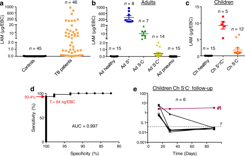
An estimated one-third of tuberculosis (TB) cases go undiagnosed or unreported. Sputum samples, widely used for TB diagnosis, are inefficient at detecting infection in children and paucibacillary patients. Indeed, developing point-of-care biomarker-based diagnostics that are not sputum-based is a major priority for the WHO. Here, in a proof-of-concept study, we tested whether pulmonary TB can be detected by analyzing patient exhaled breath condensate (EBC) samples. We find that the presence of Mycobacterium tuberculosis (Mtb)-specific lipids, lipoarabinomannan lipoglycan, and proteins in EBCs can efficiently differentiate baseline TB patients from controls. We used EBCs to track the longitudinal effects of antibiotic treatment in pediatric TB patients. In addition, Mtb lipoarabinomannan and lipids were structurally distinct in EBCs compared to ex vivo cultured bacteria, revealing specific metabolic and biochemical states of Mtb in the human lung. This provides essential information for the rational development or improvement of diagnostic antibodies, vaccines and therapeutic drugs. Our data collectively indicate that EBC analysis can potentially facilitate clinical diagnosis of TB across patient populations and monitor treatment efficacy. This affordable, rapid and non-invasive approach seems superior to sputum assays and has the potential to be implemented at point-of-care.


Conflict of interest statement

The authors declare no competing interests.

Figures

Fig. 1
Fig. 1. Quantification of LAM in EBC from TB patients and control individuals listed in Table 1.
Quantity of LAM in EBC samples from all the subjects involved (a), adults (b), and children (c, e) was determined by an immunoassay using the CS-35 anti-LAM antibody. A receiver-operating characteristic (ROC) analysis of the LAM quantitation data is shown in d. AUC area under the curve, T threshold. In a, b, and c, the difference between TB patient groups and controls (healthy, pneumo) was statistically significant (Mann–Whitney U-test, two-tailed). Error bars represent SEM. Source data are provided as a Source Data file.
Fig. 2
Fig. 2. Characterization of LAM in EBC by NMR.
Expanded region (δ 1H: 4.80-5.50, δ 13C 98-114) of the 2D 1H-13C HSQC spectrum in D2O at 298 K of Ad S+ pool (a) and Ch S+/C+ pool (b) LAM-enriched fractions, and Mtb_broth LAM (c). Cartoons show the structure of arabinan side chain termini deducted from NMR data. The branched hexa-arabinofuranoside (Ara6) motif is the main epitope of the CS-35 anti-LAM antibody. Structural motifs that differ between LAM in EBC and LAM purified from M. tuberculosis H37Rv grown in broth are highlighted in blue.
Fig. 3
Fig. 3. M. tuberculosis lipids and corresponding MS signatures detected in EBC from TB patients.
a Negative MALDI-TOF mass spectrum of PI and PIMs. A structure of tetra-acylated PIM2 (Ac2PIM2) that contains 2 palmitic (C16), 1 stearic (C18) and 1 tuberculostearic (C19) acids is drawn. * indicate intense ions that do not correspond to PIM molecular species. b Negative MALDI-TOF mass spectrum of Ac4SGL. A structure that contains 2 hydroxyphthioceranyl (HPA) and 1 phthioceranyl (PA) (SL-II according to the nomenclature of Goren) is drawn. c Positive ESI-QTOF mass spectrum of PDIM. MCA, mycocerosic acid. d, e Negative ESI-QTOF mass spectrum of α- (d) and methoxy-(e) mycolic acids. The main forms are illustrated. f Positive ESI-QTOF mass spectrum of TbAd. 1-TbAd isomer is shown. Data are representative of at least 2 independent experiments on each EBC pooled sample. The precise stereochemistry of PIMs, SGLs, PDIM and Mycolic acids can be found in Minnikin & Brennan, 2020. A detailed peak assignment is shown in Supplementary Tables 4–7.
Fig. 4
Fig. 4. Abundance of M. tuberculosis lipids in EBC from TB patients and control individuals listed in Table 1.
Abundance of MA (ac), TbAd (df), and PDIM (gi) per EBC from adults and children was determined by SFC-HRMS relatively to 1,2-ditridecanoyl-sn-glycero-3-phosphocholine (133 ng/mL of EBC) used as an internal standard (IS). Values are given as the ratio between areas of the extracted ion chromatograms (AEIC) of the ionized lipid molecular species and AEIC of the IS. In g, h, and i, values are multiplied by 10. In a, b, d, e, g, h, unless otherwise stated (ns, not significant), the difference between TB patient groups and controls (healthy, pneumo) was statistically significant (Mann–Whitney U-test, two-tailed). Error bars represent SEM. Source data are provided as a Source Data file.
Fig. 5
Fig. 5. Abundance of M. tuberculosis proteins in EBC from TB patients and control individuals listed in Table 1.
a Number of Mtb proteins detected by proteomic analysis in the corresponding groups. bi Abundance of selected Mtb proteins by proteomic analysis. jl Abundance of GroEL2 protein by an immunoassay. In bi, values are given as the Label-Free Quantification (LFQ) intensity (int.). The noise background intensity was ~3.3 log. In jl, values are given in arbitrary units corresponding to intensity (int.) on the Dot Blot (DB) and normalized to levels of GroEL2 in the Mtb cell lysate. In j and k, unless otherwise stated (ns, not significant), the difference between TB patient groups and controls (healthy, pneumo) was statistically significant (Mann–Whitney U-test, two-tailed). Error bars represent SEM. Source data are provided as a Source Data file.

References

    1. WHO. Global Tuberculosis Report 2021. 1–43 (WHO, 2021).
    1. WHO. Global Strategy and Targets for Tuberculosis Prevention, Care and Control after 2015. 1–3 (WHO, 2014).
    1. WHO. High-priority Target Product Profiles for New Tuberculosis Diagnostics: Report of A Consensus Meeting. 1–97 (WHO, 2014).
    1. Kik SV, Denkinger CM, Casenghi M, Vadnais C, Pai M. Tuberculosis diagnostics: which target product profiles should be prioritised? Eur. Respir. J. 2014;44:537–540. doi: 10.1183/09031936.00027714. - DOI - PubMed
    1. Bulterys MA, et al. Point-of-care urine LAM tests for tuberculosis diagnosis: a status update. J. Clin. Med. 2019;9:111. doi: 10.3390/jcm9010111. - DOI - PMC - PubMed

Publication types