. 2022 Dec 14;10(1):180.
doi: 10.1186/s40478-022-01480-y.

SNCA genetic lowering reveals differential cognitive function of alpha-synuclein dependent on sex

Affiliations

SNCA genetic lowering reveals differential cognitive function of alpha-synuclein dependent on sex

Jennifer L Brown et al. Acta Neuropathol Commun. .

Erratum in

Abstract

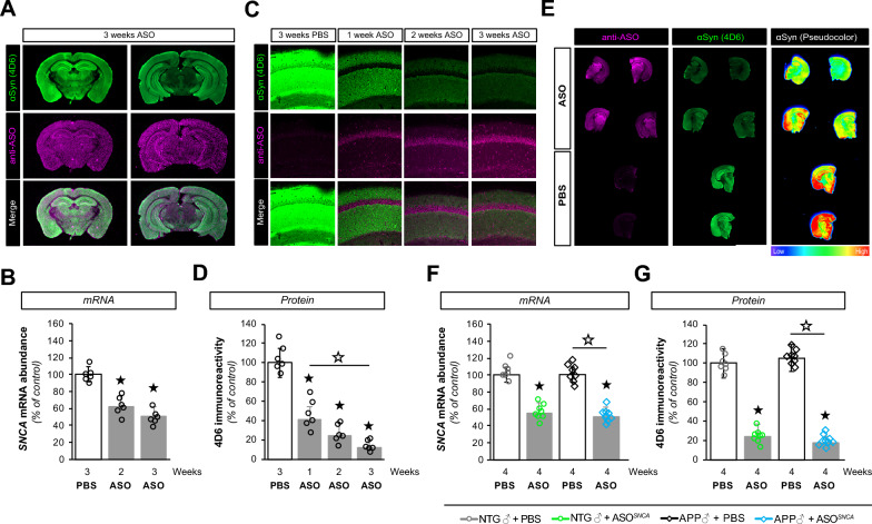
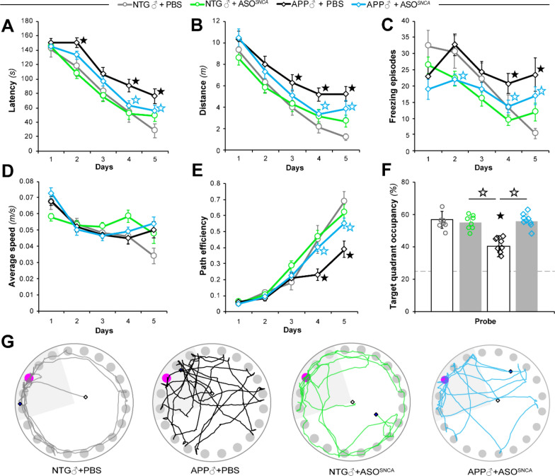
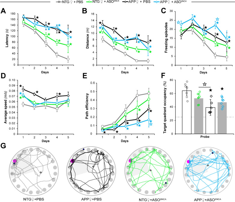
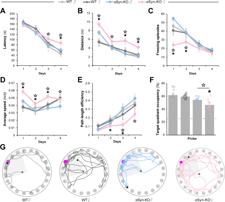
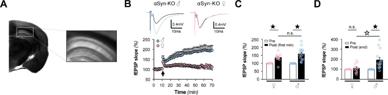
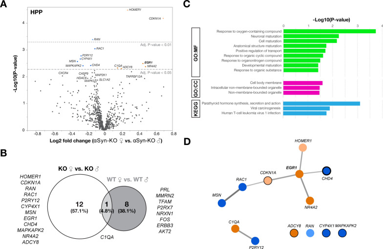
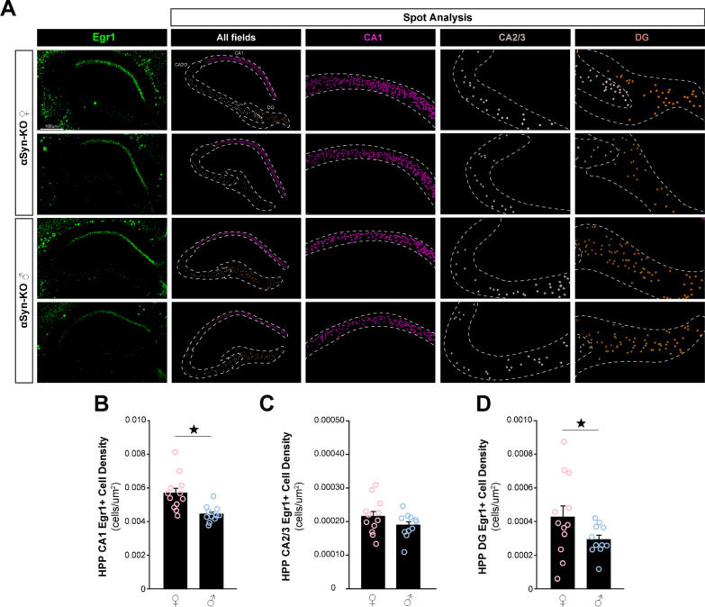
Antisense oligonucleotide (ASO) therapy for neurological disease has been successful in clinical settings and its potential has generated hope for Alzheimer's disease (AD). We previously described that ablating SNCA encoding for α-synuclein (αSyn) in a mouse model of AD was beneficial. Here, we sought to demonstrate whether transient reduction of αSyn expression using ASOSNCA could be therapeutic in a mouse model of AD. The efficacy of the ASOSNCA was measured via immunocytochemistry, RT-qPCR and western blotting. To assess spatial learning and memory, ASOSNCA or PBS-injected APP and non-transgenic (NTG) mice, and separate groups of SNCA-null mice, were tested on the Barnes circular maze. Hippocampal slice electrophysiology and transcriptomic profiling were used to explore synaptic function and differential gene expression between groups. Reduction of SNCA transcripts alleviated cognitive deficits in male transgenic animals, but surprisingly, not in females. To determine the functional cause of this differential effect, we assessed memory function in SNCA-null mice. Learning and memory were intact in male mice but impaired in female animals, revealing that the role of αSyn on cognitive function is sex-specific. Transcriptional analyses identified a differentially expressed gene network centered around EGR1, a central modulator of learning and memory, in the hippocampi of SNCA-null mice. Thus, these novel results demonstrate that the function of αSyn on memory differs between male and female brains.

Keywords: Alpha-synuclein; Alzheimer’s disease; Antisense oligonucleotide; Early growth response 1; Sex; Spatial memory; Synucleinopathy.


Conflict of interest statement

Unless stated subsequently, the authors declare that they have no competing interests. T. Cole: Other Research Support (receipt of drugs, supplies, equipment or other in-kind support); Provided drug (mouse alpha-synuclein ASO).

Figures

Fig. 1
Fig. 1
ASO1 disperses throughout the brain and lowers SNCA gene expression. A Infra-red imaging documenting the widespread distribution of ASOs 3 weeks after injection using anti- ASO (pink) and anti-αSyn (4D6, green) antibodies. B Reduction in SNCA mRNA at 2 and 3 weeks post-injection as determined by RT-qPCR. C Confocal imaging illustrating the presence of ASO1 (pink) and a corresponding decrease in αSyn (green) in mouse hippocampi. D Quantification of hippocampal 4D6 immunoreactivity in ASO1 or PBS treated mice. E Infra-red imaging detected αSyn (green) and ASO (pink) in coronal brain sections from PBS and ASO treated mice. The relative αSyn signal was lower in ASO-injected animals than in PBS-injected animals (pseudocolor). F, G Measurements of SNCA mRNA abundance by RT-qPCR (F) and αSyn protein amounts by immunofluorescence (G) in transgenic (APP) and non-transgenic (NTG) animals treated with PBS or ASO. Histogram bars represent mean ± SEM, ★P < 0.05 compared to NTG + PBS, ☆P < 0.05 compared to APP + PBS
Fig. 2
Fig. 2
Behavioral effects of αSyn reduction in male NTG and APP mice. Male APP and NTG controls were tested in the Barnes circular maze (BCM) at six months of age. A, B Spatial learning of the BCM task was reflected by reductions in escape latency (A) and distance traveled (B). C, D Measurements of freezing episode numbers (C) and average animal speed (D) in the BCM task were used to assess phenotypic changes other than spatial learning. E Quantitative assessment of animal trajectories during the learning phase using path efficiency. F, G Spatial memory retention in the BCM task was determined by target quadrant occupancy (F) and by assessment of path traces (G). Data represent mean ± SEM; ★P < 0.05 compared to NTG + PBS, ☆P < 0.05 compared to mice of the same genotype in the other treatment group, n = 8 mice/treatment/genotype
Fig. 3
Fig. 3
Behavioral effects of αSyn reduction in female NTG and APP mice. Female APP and NTG controls were tested in the Barnes circular maze (BCM) at six months of age. A, B Spatial learning of the BCM task was reflected by reductions in escape latency (A) and distance traveled (B). C, D Measurements of freezing episode numbers (C) and average animal speed (D) in the BCM task were used to assess phenotypic changes other than spatial learning. (E) Quantitative assessment of trajectories used by animals during the learning phase using path efficiency. F, G Spatial memory retention in the BCM task was determined by target quadrant occupancy (F) and by assessment of path traces (G). Data represent mean ± SEM; ★P < 0.05 compared to NTG + PBS, ☆P < 0.05 compared to mice of the same genotype in the other treatment group, n = 8 mice/treatment/genotype
Fig. 4
Fig. 4
Genetic ablation of αSyn negatively impacts the performance of female mice on the Barnes circular maze. Male and female αSyn knock-out mice (αSyn-KO) and wild-type C57BL/6 J controls were tested in the Barnes circular maze (BCM) at six months of age. A, B Spatial learning of the BCM task was reflected by reductions in escape latency (A) and distance traveled (B). C, D Measurements of freezing episode numbers (C) and average animal speed (D) in the BCM task were used to assess phenotypic changes other than spatial learning. (E) Quantitative assessment of trajectories used by animals during the learning phase using path efficiency. (F-G) Spatial memory retention in the BCM task was determined by target quadrant occupancy (F) and by assessment of path traces (G). Data represent mean ± SEM; ★P < 0.05 compared to WT mice of the same sex, ☆P < 0.05 compared to mice of the same genotype and opposite sex, n = 8 mice/genotype/sex
Fig. 5
Fig. 5
Sex differences in αSyn-KO mice hippocampal LTP. A Field excitatory postsynaptic potentials (fEPSPs) were recorded at CA1 synapses by stimulating the Schaffer collaterals. B Top, Representative fEPSP traces before (black) and after a high-frequency stimulation (HFS) of the Schaffer collaterals recorded from male (blue) and female (pink) αSyn-KO mice. Bottom, fEPSPs recorded from hippocampal CA1 during long-term potentiation induced by HFS of Schaffer collaterals (100 Hz, 1 s) of αSyn-KO females (n = 12, pink) and males (n = 14, blue). Arrowhead indicates application of HFS. C, D fEPSP slopes 1 min (C) and 60 min (D) following HFS (post), relative to baseline established prior to HFS (pre), from female and male αSyn-KO mice. Data are expressed as mean ± SEM; ★P < 0.05 compared to pre-HFS within the same sex group, ☆P < 0.05 compared to mice to opposite sex
Fig. 6
Fig. 6
Differential gene expression in male and female αSyn-KO mice. NanoString® transcriptional profiling was performed on hippocampal RNA from 6-month-old female and male αSyn-KO mice. A Volcano plot of the differentially expressed genes of αSyn-KO females compared to αSyn-KO males at adjusted significance thresholds of P < 0.05 and P < 0.01. Orange indicates upregulated genes, blue indicates downregulated genes. B Venn diagram of differentially expressed genes shows little overlap between WT and αSyn-KO females and males. C Top results for GO and KEGG databases using the female and male αSyn-KO differentially expressed gene list performed with g:Profiler for functional enrichment analysis. D MCL clustering and visualization of Cytoscape clusters was performed using clusterMaker2 based on co-expression data. Analysis revealed EGR1 as a central node in the network as well as several EGR1 target genes (black circles, thickness reflects the number of responsive elements)
Fig. 7
Fig. 7
Egr1 protein expression in the hippocampus of αSyn-KO mice. Immunofluorescence labeling and confocal microscopy image analysis of hippocampal slices from male and female αSyn-KO mice were performed for Egr1 density. A Representative 20× confocal images for Egr1 (green) and corresponding segmentations using Imaris spot analysis (CA1, magenta; CA2/3, grey; DG; brown). Hippocampal regions are indicated by dashed white lines while Egr1-positive neurons are indicated by spots. C, D Cell density analysis of Egr1-expressing neurons in CA1 (B), CA2/3 (C) and dentate gyrus (DG; D). Histogram bars represent mean ± SEM, ★P < 0.05, n = 6 mice/sex, 2 slices/mouse

References

    1. Abeliovich A, Schmitz Y, Fariñas I, Choi-Lundberg D, Ho WH, Castillo PE, Shinsky N, Garcia Verdugo JM, Armanini M, Ryan A, Hynes M, Phillips H, Sulzer D, Rosenthal A. Mice lacking α-synuclein display functional deficits in the nigrostriatal dopamine system. Neuron. 2000;25:239–252. doi: 10.1016/S0896-6273(00)80886-7. - DOI - PubMed
    1. Alarcón-Arís D, Pavia-Collado R, Miquel-Rio L, Coppola-Segovia V, Ferrés-Coy A, Ruiz-Bronchal E, Galofré M, Paz V, Campa L, Revilla R, Montefeltro A, Kordower JH, Vila M, Artigas F, Bortolozzi A. Anti-α-synuclein ASO delivered to monoamine neurons prevents α-synuclein accumulation in a Parkinson’s disease-like mouse model and in monkeys. EBioMedicine. 2020;59:102944. doi: 10.1016/J.EBIOM.2020.102944. - DOI - PMC - PubMed
    1. Bachhuber T, Katzmarski N, McCarter JF, Loreth D, Tahirovic S, Kamp F, Abou-Ajram C, Nuscher B, Serrano-Pozo A, Müller A, Prinz M, Steiner H, Hyman BT, Haass C, Meyer-Luehmann M. Inhibition of amyloid-β plaque formation by α-synuclein. Nat Med. 2015;21:802–807. doi: 10.1038/nm.3885. - DOI - PubMed
    1. Bakalash S, Pham M, Koronyo Y, Salumbides BC, Kramerov A, Seidenberg H, Berel D, Black KL, Koronyo-Hamaoui M. Egr1 expression is induced following glatiramer acetate immunotherapy in rodent models of glaucoma and alzheimer’s disease. Invest Ophthalmol Vis Sci. 2011;52:9033–9046. doi: 10.1167/IOVS.11-7498. - DOI - PubMed
    1. Bennett CF, Krainer AR, Cleveland DW. Antisense oligonucleotide therapies for neurodegenerative diseases. Annu Rev Neurosci. 2019;42:385–406. doi: 10.1146/annurev-neuro-070918-050501. - DOI - PMC - PubMed

Publication types