. 2022 Dec 1;15(12):dmm049471.
doi: 10.1242/dmm.049471. Epub 2022 Dec 15.

An induced pluripotent stem cell-based model identifies molecular targets of vincristine neurotoxicity

Affiliations

An induced pluripotent stem cell-based model identifies molecular targets of vincristine neurotoxicity

Neng-Wei Tsai et al. Dis Model Mech. .

Abstract

To model peripheral nerve degeneration and investigate molecular mechanisms of neurodegeneration, we established a cell system of induced pluripotent stem cell (iPSC)-derived sensory neurons exposed to vincristine, a drug that frequently causes chemotherapy-induced peripheral neuropathy. Sensory neurons differentiated from iPSCs exhibit distinct neurochemical patterns according to the immunocytochemical phenotypes, and gene expression of peripherin (PRPH, hereafter referred to as Peri) and neurofilament heavy chain (NEFH, hereafter referred to as NF). The majority of iPSC-derived sensory neurons were PRPH positive/NEFH negative, i.e. Peri(+)/NF(-) neurons, whose somata were smaller than those of Peri(+)/NF(+) neurons. On exposure to vincristine, projections from the cell body of a neuron, i.e. neurites, were degenerated quicker than somata, the lethal concentration to kill 50% (LC50) of neurites being below the LC50 for somata, consistent with the clinical pattern of length-dependent neuropathy. We then examined the molecular expression in the MAP kinase signaling pathways of, extracellular signal-regulated kinases 1/2 (MAPK1/3, hereafter referred to as ERK), p38 mitogen-activated protein kinases (MAPK11/12/13/14, hereafter referred to as p38) and c-Jun N-terminal kinases (MAPK8/9/10, hereafter referred to as JNK). Regarding these three cascades, only phosphorylation of JNK was upregulated but not that of p38 or ERK1/2. Furthermore, vincristine-treatment resulted in impaired autophagy and reduced autophagic flux. Rapamycin-treatment reversed the effect of impaired autophagy and JNK activation. These results not only established a platform to study peripheral degeneration of human neurons but also provide molecular mechanisms for neurodegeneration with the potential for therapeutic targets.

Keywords: Autophagy; IPSC; MAP kinase; Nerve degeneration; Sensory neuron; Vincristine.


Conflict of interest statement

Competing interests The authors declare no competing or financial interests.

Figures
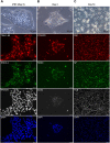
Fig. 1.
Fig. 1.
The morphology and protein expression patterns of iPSC-derived sensory neurons during differentiation process. Top three images: Cell morphology was observed at days 0, 3 and 14 of neuronal differentiation using bright field microscopy; scale bars: 50 µm. All other images: Differential stage markers were examined by using immunocytochemistry; scale bars: 20 µm. (A) Images of cells stained for TRA-1-60, SSEA-4 and NANOG on day 0. (B) Images of cells expressing the neural stem cell markers vimentin, nestin and SOX2 on day 3. (C) Images of differentiated neurons expressing the cytoskeletal proteins peripherin (Peri), neurofilament (NF) and βIII-tubulin (TUB) on day 14. DAPI was used to stain nuclei.
Fig. 2.
Fig. 2.
Temporal patterns of gene expression during neuronal differentiation. mRNA expression levels during the stages of differentiation (days 0-15) were examined using qPCR. (A) Pluripotency genes NANOG, OCT4 and REX1. (B) Genes encoding neural stem cell marker SOX2, neural progenitor cell marker PAX6 and factor of participated neuronal differentiation ASCL1. (C) Neuron-specific genes: PRPH, TUJ1 and TRPV1. (D,E) Compared are expression levels of TRK receptors TRKA, TRKB and TRKC (D), and expression levels of transcription factors RUNX1, SHOX2 and RUNX3 (E) to distinguish between the subtype of iPSC-derived sensory neurons. mRNA expression levels are plotted as ΔCt, i.e. the difference between the Ct value of each gene in question and that of the GAPDH (internal control). Levels are shown relative to those on day 0. One-way ANOVA followed by Tukey's multiple comparisons test, *P<0.05, **P<0.01 compared to day 0; n=3.
Fig. 3.
Fig. 3.
Soma size of Peri(+)/NF(+) neurons and Peri(+)/NF(−) neurons. iPSC-derived sensory neurons, i.e. Peri(+)/NF(+) and Peri(+)/NF(−) neurons, were stained against peripherin (Peri) and neurofilament (NF) to compare their pattern of soma size and gene expression. (A) iPSCs were differentiated into two phenotypes: Peri(+)/NF(+) (yellow) or Peri(+)/NF(−) neurons (red), scale bars: 20 µm. (B) Confocal microscopy images illustrate how neurons of both phenotype were defined. To avoid incorrectly classifying neurites as neurons, images were pre-processed by adjusting the threshold of neurite signals, i.e. neurons of both phenotypes were automatically classified by the morphometric software ImageJ, allowing measurement of soma size for each neuron. (C) Histogram showing the distribution of soma size for Peri(+)/NF(+) neurons versus Peri(+)/NF(−) neurons. Somata of Peri(+)/NF(+) neurons were larger compared with those of Peri(+)/NF(−) neurons. n=3. (D) iPSC-derived sensory neurons were sorted using a 4-laser FACSAriaIII cell sorter and gene expression patterns were examined by qPCR. Peri(+)/NF(−) neurons showed significantly higher expression of TRKA than Peri(+)/NF(+) neurons, but Peri(+)/NF(+) neurons showed significantly higher expression of NEFH (NFH) CGRP and TAC1. No significant differences were found regarding expression of RET and RUNX1. One-way ANOVA followed by Tukey's multiple comparisons test, *P<0.05 and **P<0.01 compared to Peri(+)/NF(+) neurons. n=3.
Fig. 4.
Fig. 4.
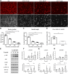
Neurotoxic effects and activation of the MAP kinase signaling pathway (JNK, p38 and ERK) during vincristine-induced neurodegeneration. iPSC-derived sensory neurons were treated with vincristine to investigate vincristine neurotoxicity and the effect of the drug on expression of JNK, p38 and ERK. (A) Neurons were treated for 48 h with different concentrations of vincristine (0.47, 1.88 or 7.5 nM). The neurotoxic effects of vincristine on soma versus neurite were assessed with immunostaining against peripherin (red) and βIII-tubulin (white). Scale bars: 20 µm. Images in this panel were used for quantification of neuron numbers and neurite lengths as shown in B and C, and to quantify the axon degeneration index (see Fig. S3B). The Ctrl and VCR 7.5 nM βIII-tubulin images are reused in Fig. S3A to illustrate the axon degradation analysis. (B,C) Quantitative analysis of neuron number and neurite length showed distinct pattern in soma versus neurite. (D) LC50 analysis, confirming that neurites are more affected by vincristine than somata. (E-H) iPSC-derived sensory neurons were treated with 7.5 nM vincristine for 0, 12, 24 and 48 h. Protein levels of JNK, p38 and ERK1/2 and their phosphorylated versions (p-JNK, p-p38, p-ERK, respectively) was assessed by western blotting; β-actin was used as an internal control. Quantification indicated a time-dependent increase after vincristine treatment only for p-JNK. One-way ANOVA followed by Tukey's multiple comparisons test, *P<0.05 and **P<0.01 compared to untreated cells, n=3.
Fig. 5.
Fig. 5.
The effect of vincristine on autophagy. Effect of vincristine on protein levels of the autophagy-related molecules membrane-bound lipidated MAP1LC3A and SQSTM1 (LC3-II and p62, respectively) were examined with western blotting. iPSC-derived sensory neurons were treated for 24 h with 7.5 nM vincristine, the autophagy inducer rapamycin (200 nM) or the autophagy blocker bafilomycin A1 (10 nM), or were left untreated (−), and levels of cytoplasmic MAP1LC3A (LC3-I), LC3-II and p62 were compared. (A) Western blot showing that LC3-II and p62 protein levels were increased after vincristine and bafilomycin A1 treatment compared with levels in the untreated group (control group). (B,C) Quantitative analysis of the western blotting experiment. Normalization to levels of β-actin confirmed the increase of LC-II (A) and p62 (B) in vincristine- and bafilomycin A1-treated cells, indicating that autophagy was impaired after vincristine treatment. One-way ANOVA followed by Tukey's multiple comparisons test, *P<0.05 and **P<0.01 compared to untreated group (control group), n=4.
Fig. 6.
Fig. 6.
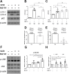
Effects of vincristine on autophagy and the MAP kinase signaling pathway. iPSC-derived sensory neurons were treated with vincristine to assess the relationship between autophagy and JNK protein levels. (A-C) Degradation of LC3 and p62 was determined in a turnover assay by adding bafilomycin A1 (Baf A1). Neurons were treated with 7.5 nM vincristine in the presence and absence of 10 nM bafilomycin A1 for 12 h. Following this, the differences in LC3 or p62 densitometric levels (DV) of the vincristine-treated groups – i.e. (④-③) or (⑧-⑦), respectively – were compared with that of the matching control groups – i.e. (②-①) or (⑥-⑤), respectively. (D,E) Differences in LC3-II and p62 protein levels in the presence or absence of bafilomycin A1 were normalized to protein levels in untreated neurons (control group), and are shown as relative degeneration levels. Relative degradation of p62 was markedly reduced after vincristine treatment. (F-H) Levels of phosphorylated JNK (p-JNK) were examined by western blotting; β-actin was used as an internal control. In the presence of bafilomycin A1, p-JNK levels in vincristine-treated sensory neurons were higher compared with those without bafilomycin A1. One-way ANOVA followed by Tukey's multiple comparisons test, *P<0.05 and **P<0.01 compared to untreated (control) cells, n=4.
Fig. 7.
Fig. 7.
Effect of rapamycin on vincristine-induced autophagy impairment in the presence or absence of bafilomycin A1. iPSC-derived sensory neurons were pretreated with different concentrations (100 nM, 200 nM or 400 nM) of rapamycin for 24 h and then treated with 7.5 nM vincristine in the presence or absence of 10 nM bafilomycin A1 for 24 h. (A) Autophagy-related molecules (LC3-II and p62) were examined by western blotting with β-actin as internal control. (B) Western blot results were quantified with densitometry. Differences between neurons treated with or without bafilomycin A1 were normalized to those in the untreated rapamycin group. The results indicate that pretreatment with 200 nM or 400 nM rapamycin can reverse vincristine-induced autophagy. Paired t-test, *P<0.05, n=4.
Fig. 8.
Fig. 8.
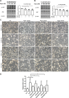
Effects of rapamycin on vincristine-induced JNK phosphorylation. (A,B) iPSC-derived sensory neurons were pretreated with various concentrations (100 nM, 200 nM, 400 nM) of rapamycin for 24 h, followed by treatment with 7.5 nM vincristine (VCR) for 24 h (A) and 48 h (B). Levels of phosphorylated JNK (p-JNK) were examined by western blotting; β-actin was used as an internal control, and quantified using densitometry. These results suggest that JNK phosphorylation induced by treatment with vincristine for 24 h and 48 h is significantly reduced following pretreatment with 200 nM or 400 nM rapamycin. Paired t-test, *P<0.05 and **P<0.01, n=5. (C) Bright field images, showing no difference in neuron number after pretreatment with rapamycin. Pretreatment with DMSO was used a control. (D) Degeneration index analysis, showing no difference in the neurite degeneration index after pretreatment with rapamycin at different concentrations followed by treatment with 7.5 nM vincristine for 48 h. Paired t-test, *P<0.05 and **P<0.01, n=6.

References

    1. Ahmed, T., Zulfiqar, A., Arguelles, S., Rasekhian, M., Nabavi, S. F., Silva, A. S. and Nabavi, S. M. (2020). Map kinase signaling as therapeutic target for neurodegeneration. Pharmacol. Res. 160, 105090. 10.1016/j.phrs.2020.105090 - DOI - PubMed
    1. Bekker, M., Abrahams, S., Loos, B. and Bardien, S. (2021). Can the interplay between autophagy and apoptosis be targeted as a novel therapy for Parkinson's disease? Neurobiol. Aging 100, 91-105. 10.1016/j.neurobiolaging.2020.12.013 - DOI - PubMed
    1. Bianchi, F., Malboubi, M., Li, Y., George, J. H., Jerusalem, A., Szele, F., Thompson, M. S. and Ye, H. (2018). Rapid and efficient differentiation of functional motor neurons from human iPSC for neural injury modelling. Stem Cell Res 32, 126-134. 10.1016/j.scr.2018.09.006 - DOI - PubMed
    1. Bráz, J. M. and Basbaum, A. I. (2010). Differential ATF3 expression in dorsal root ganglion neurons reveals the profile of primary afferents engaged by diverse noxious chemical stimuli. Pain 150, 290-301. 10.1016/j.pain.2010.05.005 - DOI - PMC - PubMed
    1. Bruna, J., Alberti, P., Calls-Cobos, A., Caillaud, M., Damaj, M. I. and Navarro, X. (2020). Methods for in vivo studies in rodents of chemotherapy induced peripheral neuropathy. Exp. Neurol. 325, 113154. 10.1016/j.expneurol.2019.113154 - DOI - PMC - PubMed

Publication types

MeSH terms

Substances