

Multiscale quantification of tumor microarchitecture for predicting therapy response using dynamic contrast-enhanced ultrasound imaging
- PMID: 36518354
- PMCID: PMC9745672
- DOI: 10.1109/ultsym.2019.8926152
Multiscale quantification of tumor microarchitecture for predicting therapy response using dynamic contrast-enhanced ultrasound imaging
Abstract
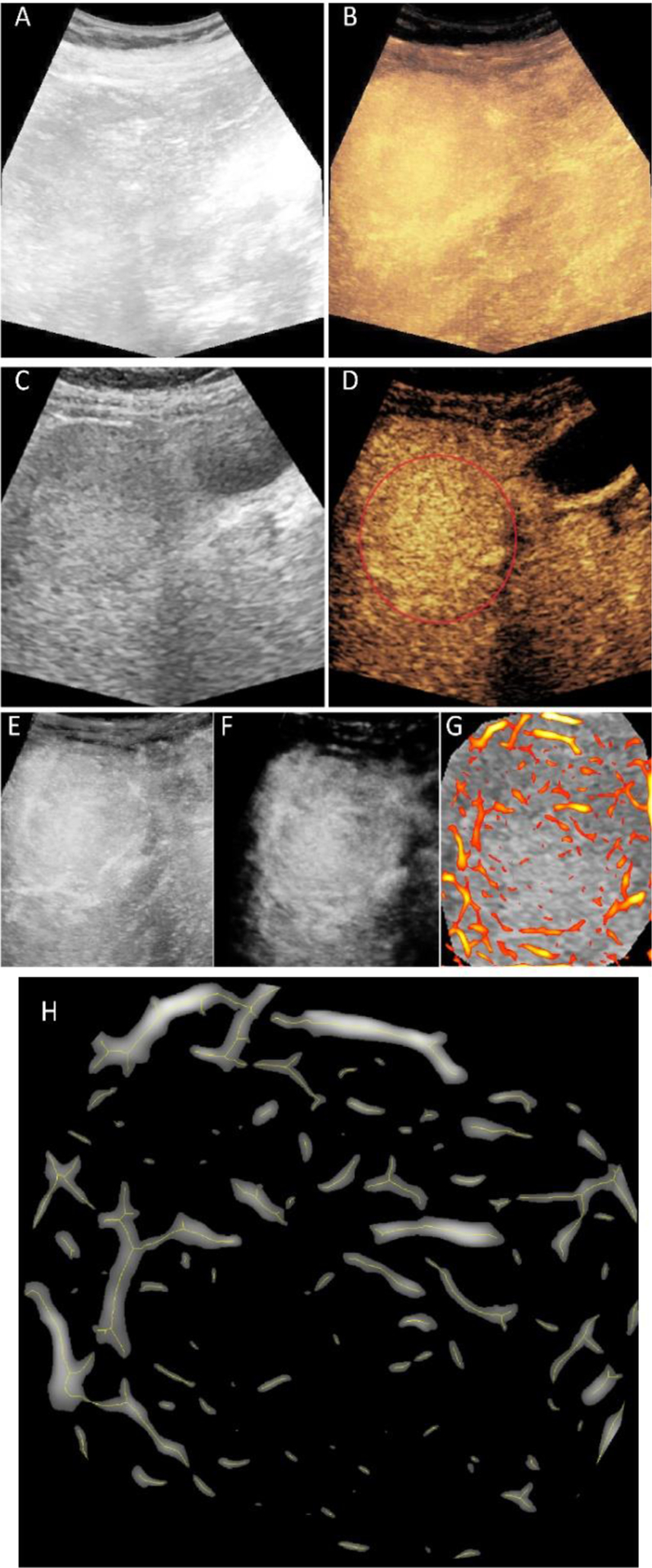
Hepatocellular carcinoma (HCC) is the most common liver cancer with 1 million cases globally. A current clinical challenge is to determine which patients will respond to transarterial chemoembolization (TACE) as effective delivery of the embolic material may be influenced by the tumor vascular supply. The purpose of this study is to develop a novel image processing algorithm for improved quantification of tumor microvascular morphology features using contrast-enhanced ultrasound (CEUS) images and to predict the TACE response based on these biomarkers before treatment. A temporal sequence of CEUS images was corrected from rigid and non-rigid motion artifacts using affine and free form deformation models. Subsequently, a principal component analysis based singular value filter was applied to remove the clutter signal from each frame. A maximum intensity projection was created from high-resolution images. A multiscale vessel enhancement filter was first utilized to enhance the tubular structures as a preprocessing step before segmentation. Morphological image processing methods are used to extract the morphology features, namely, number of vessels (NV) and branching points (NB), vessel-to-tissue ratio (VR), and the mean vessel length (VL), tortuosity (VT), and diameter (VD) from the tumor vascular network. Finally, a support vector machine (SVM) is trained and validated using leave-one-out cross-validation technique. The proposed image analysis strategy was able to predict the patient outcome with 90% accuracy when the SVM was trained with the three features together (NB, NV, VR). Experimental results indicated that morphological features of tumor microvascular networks may be significant predictors for TACE response. Reliable prediction of the TACE therapy response may help provide effective therapy planning.
Keywords: cancer; contrast-enhanced ultrasound; hepatocellular carcinoma; image analysis; machine learning; microbubble; microvascular networks; transarterial chemoembolization.
Figures
References
-
- Shaw CM et al., “Contrast-Enhanced Ultrasound Evaluation of Residual Blood Flow to Hepatocellular Carcinoma After Treatment With Transarterial Chemoembolization Using Drug-Eluting Beads,” Journal of Ultrasound in Medicine, vol. 34, no. 5, pp. 859–867, 2015. - PubMed
Grants and funding
LinkOut - more resources
Full Text Sources
Other Literature Sources
Miscellaneous