. 2022 Oct 17:9:1920-1928.
doi: 10.1016/j.toxrep.2022.10.006. eCollection 2022.

In vitro and in vivo hepatotoxicity study of Afriplex™ GRT through an inflammatory response

Affiliations

In vitro and in vivo hepatotoxicity study of Afriplex™ GRT through an inflammatory response

Kwazikwakhe B Gabuza et al. Toxicol Rep. .

Abstract

Background: The focus on traditional and complementary medicine for supplementation and treatment of diseases is high. Aspalathus linearis commonly known as Rooibos showed several beneficial effects, this led to the standardized production of a pharmaceutical grade green rooibos extract (Afriplex TM GRT) with enhanced polyphenolic content. The aim of this study was to assess toxicity of Afriplex TM GRT in HepG2/C3A cells and Sprague Dawley rats.

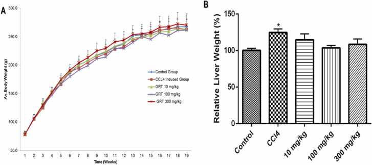
Methods: Afriplex GRT TM (0.1, 1, 10, 100, or 1000 μg/mL) in DMSO was added to the media to the final 0.01% DMSO for treatment of HepG2/C3A for 1, 24 and 48 hrs followed by MTT and ATP assays. Sprague Dawley rats were grouped to Control, Afriplex TM GRT treated (10, 100 and 300 mg/kg); and acute (24hrs tetrachloromethane (CCl 4) injected hepatotoxicity control). Serum biochemistry, histology and Western blot analysis on liver were performed.

Results: Afriplex TM GRT significantly reduced cell viability at 100 and 1000μg/mL after 48 hrs. Acute CCl 4 treatment significantly increased serum alanine aminotransferase in rats. The highest extract treatment of 300 mg/kg significantly elevated aspartate amino transferase. There was severe macro vesicular in the CCl 4 group whereas mild to moderate micro vesicular steatosis was seen in the 300 mg/kg Afriplex TM GRT treated group. Highest extract treatment significantly reduced NFkB expression on Western blot analysis.

Conclusion: The beneficial effects of pharmaceutical grade Afriplex GRT TM are concentration and dosage based. Afriplex GRT TM exerts its beneficial effects via NFkB as demonstrated by the dose dependent reduction of NFkB on Western blot analysis. More work need to be done to explore the exact mechanism that occurs in the NFkB pathway.

Keywords: Aspalathin-rich; HepG2/C3A; Hepatotoxicity; Rooibos; Sprague Dawley.


Conflict of interest statement

The authors declare that they have no known competing financial interests or personal relationships that could have appeared to influence the work reported in this paper.

Figures

ga1
Graphical abstract
Fig. 1
Fig. 1
The effect of Afriplex™ GRT on cell viability on HepG2/C3A cells using MTT and ATP assays. The effect of Afriplex™ GRT on HepG2/C3A cells viability after 1 h (A), 24 h (B), and 48 h (C) incubation using MTT assay. Cell viability was confirmed by ATP assay after 1 h (D), 24 h (E), and 48 h (F) incubation. The data is presented as mean ± SEM of three independent experiments, * ** p-value < 0.001 versus control, *p < 0.05.
Fig. 2
Fig. 2
The effect of Afriplex™ GRT on body weight (A) and relative liver weight (B) of Sprague-Dawley rats. The body weights were monitored weekly and recorded as mean ± SEM and relative liver weight recorded at termination and calculated by dividing the body weight of the rat with the liver weight expressed relative to the control set at 100%, presented as Mean ± SEM. (n = 10 rats/group).
Fig. 3
Fig. 3
Liver function ALT (A), AST (B), ALP (C), total protein (D), albumin (E), total bilirubin (F), urea (G), uric acid (H) and creatinine (I) levels for the untreated control group and treated with CCl4 administered 24 h before terminations (0.8 mg/kg; positive control) and Afriplex™ GRT administered for 90 days (10, 100, 300 mg/kg) treated groups (n = 10 rats/group). Results represent the mean ± SEM. Statistical analysis One-way ANOVA, *p < 0.05 versus the vehicle control group (M), * *p < 0.01 versus vehicle control (M), #p < 0.05 versus CCl4 (F), ##p < 0.01 versus CCl4 (F), ^p < 0.05 versus CCl4 (M) and ^^p < 0.01 versus CCl4 (M).
Fig. 4
Fig. 4
Representative liver hematoxylin and eosin (H&E) sections from different treatment groups. Normal liver tissue demonstrating typical liver morphology with hepatocytes and sinusoids (A); CCl4 treated positive control group (B) demonstrates severe diffuse hepatocellular micro- and macro-vesicular degeneration (black arrows) with pyknotic nuclei (red arrows) showed central vein without micro- and macro-vesicular degeneration; The 10 mg/kg treated group (C); The 100 mg/kg treated group (D) showing minor hepatocellular micro-vesicular degeneration changes present; The 300 mg/kg treated group (E) shows a focal area of hepatocellular micro-vesicular degeneration with more acinar spaces instead of macro-vesicular degeneration. H & E 200 x magnification.
Fig. 5
Fig. 5
Representative liver Glutathione S-Transferase (GST-Pi) sections from different treatment groups. The vehicle untreated group (A). The 10 mg/kg treated group (B). The 100 mg/kg treated group (C). The 300 mg/kg treated group (D). CCl4 treated positive control group (E). GST-Pi x 200 magnification.
Fig. 6
Fig. 6
Relative expression of nuclear factor-kB (A), CYP3A4 (B), Caspase 3 (C) and Bax (D) and Bcl-2 (E)assessed by Western blot in liver of male rats (n = 5 rats /group), NF-kB p65, Bax, Caspase 3, CYP3A4 and Bcl-2 primary antibodies were used on liver samples of untreated, treated with CCl4 (0.8 mg/kg; positive control) 24 h before termination or Afriplex™ GRT (10, 100, 300 mg/kg) administered for 90 days. Results represent the mean ± SEM in three independent experiments relative to the house-keeping gene, B-Actin. *p < 0.05 versus the control group.

References

    1. Bell L.N., Chalasani N. Epidemiology of idiosyncratic drug-induced liver injury. Semin Liver Dis. 2009;29:337–347. - PMC - PubMed
    1. El-Sayed A.M.H. Possible effects of natural antioxidants on experimentally induced hepatotoxicity in rats. CU Theses. 2016;0
    1. Navarro V., Senior J. Drug-related hepatotoxicity. N. Engl. J. Med. 2006;354:731–739. - PubMed
    1. Leise M.D., Poterucha J.J., Talwalkar J.A. Drug-induced liver injury. Mayo Clin. Proc. 2014;89:95–106. - PubMed
    1. Björnsson E.S. Drug-induced liver injury: an overview over the most critical compounds. Arch. Toxicol. 2015;89:327–334. - PubMed

LinkOut - more resources