Case Reports
. 2022 Dec 5:2022:9773058.
doi: 10.1155/2022/9773058. eCollection 2022.

Neuroinvasive Onchocerca lupi Infection in a Ten-Year-Old Girl

Affiliations
Case Reports

Neuroinvasive Onchocerca lupi Infection in a Ten-Year-Old Girl

Dorothy Bowers Wu et al. Case Rep Infect Dis. .

Abstract

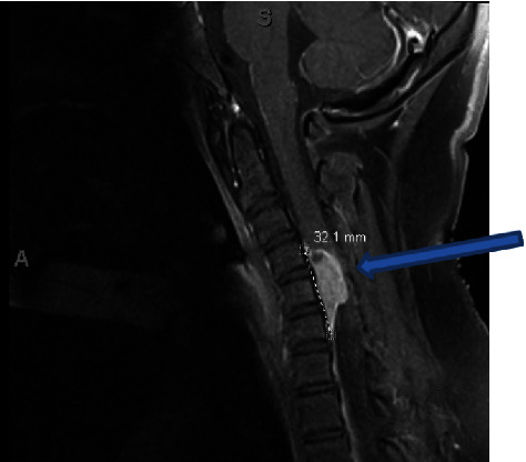
The nematode Onchocerca lupi is an emerging human pathogen. Though its life cycle is not well studied, it likely infects humans after a bite from a black fly vector, which in turn acquires infective microfilariae from an infected canid. These microfilariae mature into an infective larval stage within the fly. Among six reported cases in the United States, five involved children, and all occurred in the southwest. In this report, we present a case of O. lupi infection with cervical spine invasion in a healthy 10-year-old girl. She presented with five months of neurological symptoms from a rural and medically underserved area, highlighting a need for clinical vigilance in such settings for this emerging infectious threat in the American southwest.


Conflict of interest statement

The authors declare that they have no conflicts of interest.

Figures

Figure 1
Figure 1
Sagittal T1-weighted preoperative MRI with contrast showing an anterior intradural extramedullary mass, measuring 1.6 × 0.9 × 2.8 cm (blue arrow). The mass is centered at C5 and spans from C4-6 with what appears to be a dural tail. The mass shows significant compression of the spinal cord.
Figure 2
Figure 2
Longitudinal section revealing degenerating Onchocerca lupi nematode encased in inflamed, dense fibrous tissue. Lillie–Twort Gram stain, original magnification ×25.
Figure 3
Figure 3
Trichrome-stained sections of O. lupi revealing relevant morphological features ((a) 200x magnification and (b) 400x magnification). Arrows indicate external cuticular ridges, and arrowheads indicate internal striae.

References

    1. Cantey P. T., Weeks J., Edwards M., et al. The emergence of zoonotic Onchocerca lupi infection in the United States--A case-series. Clinical Infectious Diseases . 2016;62(6):778–783. doi: 10.1093/cid/civ983. - DOI - PMC - PubMed
    1. Rojas A., Morales-Calvo F., Salant H., Otranto D., Baneth G. Zoonotic ocular onchocercosis by Onchocerca lupi. Yale J Biol Med . 2021;94(2):331–341. - PMC - PubMed
    1. Hassan H. K., Bolcen S., Kubofcik J., et al. Isolation of Onchocerca lupi in dogs and black flies, California, USA. Emerging Infectious Diseases . 2015;21(5):789–796. doi: 10.3201/eid2105.142011. - DOI - PMC - PubMed
    1. Roe C. C., Yaglom H., Howard A., et al. Coyotes as reservoirs for Onchocerca lupi, United States, 2015 – 2018. Emerging Infectious Diseases . 2020;26(12):2989–2993. doi: 10.3201/eid2612.190136. - DOI - PMC - PubMed
    1. Eberhard M. L., Hobohm D., Chundu K., et al. Zoonotic Onchocerca lupi infection in a 22-month old child in Arizona: first report in the United States and a review of the literature. The American Journal of Tropical Medicine and Hygiene . 2013;88(3):601–605. doi: 10.4269/ajtmh.12-0733. - DOI - PMC - PubMed

Publication types

LinkOut - more resources