. 2022 Nov 28:13:1051008.
doi: 10.3389/fimmu.2022.1051008. eCollection 2022.

The immune response to lumpy skin disease virus in cattle is influenced by inoculation route

Affiliations

The immune response to lumpy skin disease virus in cattle is influenced by inoculation route

Petra C Fay et al. Front Immunol. .

Abstract

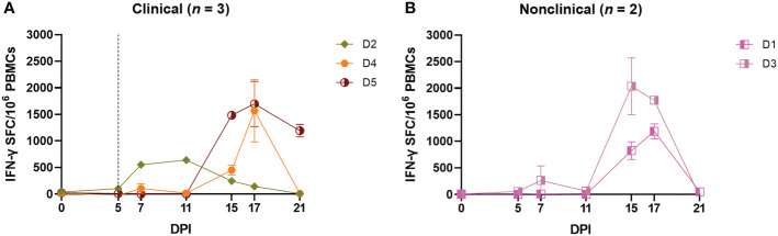
Lumpy skin disease virus (LSDV) causes severe disease in cattle and water buffalo and is transmitted by hematophagous arthropod vectors. Detailed information of the adaptive and innate immune response to LSDV is limited, hampering the development of tools to control the disease. This study provides an in-depth analysis of the immune responses of calves experimentally inoculated with LSDV via either needle-inoculation or arthropod-inoculation using virus-positive Stomoxys calcitrans and Aedes aegypti vectors. Seven out of seventeen needle-inoculated calves (41%) developed clinical disease characterised by multifocal necrotic cutaneous nodules. In comparison 8/10 (80%) of the arthropod-inoculated calves developed clinical disease. A variable LSDV-specific IFN-γ immune response was detected in the needle-inoculated calves from 5 days post inoculation (dpi) onwards, with no difference between clinical calves (developed cutaneous lesions) and nonclinical calves (did not develop cutaneous lesions). In contrast a robust and uniform cell-mediated immune response was detected in all eight clinical arthropod-inoculated calves, with little response detected in the two nonclinical arthropod-inoculated calves. Neutralising antibodies against LSDV were detected in all inoculated cattle from 5-7 dpi. Comparison of the production of anti-LSDV IgM and IgG antibodies revealed no difference between clinical and nonclinical needle-inoculated calves, however a strong IgM response was evident in the nonclinical arthropod-inoculated calves but absent in the clinical arthropod-inoculated calves. This suggests that early IgM production is a correlate of protection in LSD. This study presents the first evidence of differences in the immune response between clinical and nonclinical cattle and highlights the importance of using a relevant transmission model when studying LSD.

Keywords: Humoral immunity; Lumpy Skin Disease; bovine immunity; cell-mediated immunity; neutralising antibodies; poxvirus; virus.


Conflict of interest statement

The authors declare that the research was conducted in the absence of any commercial or financial relationships that could be construed as a potential conflict of interest.

Figures

Figure 1
Figure 1
Clinical and virological outcomes of clinical and nonclinical calves after needle–inoculation with LSDV. The number of cutaneous lesions (A, B) were recorded each day, up to a maximum of 100 lesions. The rectal temperature of clinical and nonclinical calves (C, D) was recorded daily. The number of LSDV genome copies in the blood of each calf was quantified by qPCR (E, F). A vertical dotted line represents the first day cutaneous lesions were noted in the clinical calves.
Figure 2
Figure 2
Needle–inoculated clinical and nonclinical calves produce IFN–γ but not type I IFN in response to stimulation of PBMCs or blood by UV–inactivated LSDV. (A, B) The amount of type I IFN in the sera of needle–inoculated cattle at indicated timepoints post infection was quantified using a cellular reporter system (Mx/CAT assay). IGRAs were performed on PBMC purified from heparinised blood collected at indicated timepoints from clinical (C) and nonclinical (D) calves. PBMCs were stimulated overnight with UV–inactivated LSDV, supernatant collected, and IFN–γ quantified using an in–house ELISA. The error bars represent the SEM. An IGRA was also performed on stimulated whole blood from clinical (E) and nonclinical (F) calves at the timepoints indicated. Whole blood was stimulated with live LSDV overnight, then IFN–γ present in the plasma quantified using a commercially available ELISA. The error bars represent the SEM. The dotted horizontal line represents a 15 S/P % positive cut–off. The dotted vertical line represents the first day cutaneous lesions occurred in the clinical animals. Data corrected to mock PBS stimulation.
Figure 3
Figure 3
Needle–inoculated clinical (A) and nonclinical (B) calves produce IFN–γ in response to stimulation of PBMCs by UV–inactivated LSDV as measured by ELISpot assay. The number of IFN–γ producing PBMCs stimulated with SW–UV inactivated LSDV was determined by IFN–γ ELISpot and presented as spot forming cells/million (SFC/106). The error bars represent the SEM. The dotted line represents the first day of the onset of cutaneous lesions in the clinical animals. Data corrected to mock PBS stimulation.
Figure 4
Figure 4
CD4+CD8 and CD8+CD4 but not CD4CD8 T cells are responsible for the production of IFN–γ in response to stimulation with UV–inactivated LSDV in needle–inoculated calves. Flow cytometric analysis was used to determine the % of IFN–γ+ T cells expressing CD4+CD8 (A, B), CD8+CD4 (C, D) and CD4CD8 (E, F) in response to stimulation with SW–UV inactivated LSDV. Samples were tested in triplicate and normalised to the mock control. The error bars represent the SEM. The dotted vertical line represents the first day lesions occurred in clinical animals. Data corrected to mock PBS stimulation.
Figure 5
Figure 5
Calves that develop clinical disease following needle–inoculation with LSDV also develop a more rapid and robust humoral immune response as measured either by ELISA or FVNT. The antibody responses in calves after needle–inoculation with LSDV was measured using the ID Screen® Capripox Double Antigen Multi–species ELISA kit (Innovative Diagnostics) (A, B). The horizontal line represents the 30 S/P% cut–off. The production of neutralising antibodies was measured using a fluorescent virus neutralisation test. The percentage of viral foci–forming units neutralised by the sera was measured over time in clinical (C) and nonclinical (D) calves. Complete neutralisation of the virus (FVNT100) was also calculated in clinical (E) and nonclinical (F) calves. Uninfected calves in groups A–C were negative for LSDV–specific antibodies throughout the study period. The dotted line represents the first day of the onset of cutaneous lesions in the clinical animals.
Figure 6
Figure 6
Eight out of ten arthropod–inoculated calves developed clinical signs consistent with LSD approximately 11 days after the start of inoculation. The number of cutaneous lesions in the clinical (A) and nonclinical (B) calves were recorded each day, up to a maximum of 100 lesions. The rectal temperature of clinical and nonclinical calves (C, D) was recorded daily. The number of LSDV genome copies in the blood of each calf was quantified by qPCR. (E, F) The red dotted line represents the onset of cutaneous lesions for group RS cattle and the blue dotted line the onset of cutaneous lesions for group RA cattle.
Figure 7
Figure 7
Arthropod–inoculated clinical calves but not nonclinical calves develop a uniform and robust LSDV–specific CMI response as measured by IGRA on purified PBMCs. The amount of type I IFN in the sera of needle–inoculated cattle at indicated timepoints post infection (A, B) was quantified using a cellular reporter system (MxCAT assay). IGRAs were performed on PBMC purified from heparinised blood collected at indicated timepoints from clinical (C) and nonclinical (D) calves. PBMCs were stimulated overnight with UV–inactivated LSDV, supernatant collected, and IFN–γ quantified using an in–house ELISA. The error bars represent the SEM. An IGRA was also performed on stimulated whole blood from clinical (E) and nonclinical (F) calves at the timepoints indicated. Whole blood was stimulated with live LSDV overnight, then IFN–γ present in the plasma quantified using a commercially available ELISA. The error bars represent the SEM. The dotted horizontal line represents a 15 S/P % positive cut–off. Red dotted line represents the onset of lesions for group RS cattle and the blue dotted line for group RA cattle. Data corrected to mock PBS stimulation.
Figure 8
Figure 8
Calves that develop clinical disease following arthropod inoculation with LSDV develop a rapid and robust humoral immune response as measured either by ELISA or FVNT. The antibody responses in calves after needle–inoculation with LSDV was measured using the ID Screen® Capripox Double Antigen Multi–species ELISA kit (Innovative Diagnostics) following manufacturer instructions (A, B). The error bars represent the SEM. The production of neutralising antibodies was measured using a fluorescent virus neutralisation test. The percentage of viral foci–forming units neutralised by the sera was measured over time in clinical (C) and nonclinical (D) calves. Complete neutralisation of the virus (FVNT100) was also calculated in clinical (E) and nonclinical (F) calves. Red dotted line represents the onset of lesions for group RS cattle and the blue dotted line for group RA cattle.
Figure 9
Figure 9
Protection against LSD is associated with a robust LSDV–specific IgM response following arthropod–inoculation but not needle–inoculation. PBMCs were isolated from heparinised blood from needle–inoculated (A, B) and arthropod–inoculated (C, D) calves at the time points shown, and stimulated overnight with live LSDV. Antibody secreting cells were then labelled with bovine anti–IgM or bovine anti–IgG antibodies. Spots were read using an ImmunoSpot 7.0 reader and ImmunoSpot SC suite (Cellular Technology Limited). Results were manually validated for false–positive results and expressed as the mean number of ASCs/million. Results are expressed as group mean ASCs/106 PBMCs ± standard error of the mean. Red dotted line represents the onset of lesions for group RS cattle and the blue dotted line for group RA cattle. Data corrected to mock uninfected cell lysate stimulation.

References

    1. Kitching RP, Bhat PP, Black DN. The characterization of African strains of capripoxvirus. Epidemiol Infect (1989) 102(2):335–43. doi: 10.1017/S0950268800030016 - DOI - PMC - PubMed
    1. Tuppurainen ESM, Venter EH, Shisler JL, Gari G, Mekonnen GA, Juleff N, et al. . Review: Capripoxvirus diseases: Current status and opportunities for control. Transboundary Emerging Dis (2017) 64(3):729–45. doi: 10.1111/tbed.12444 - DOI - PMC - PubMed
    1. Khan YR, Ali A, Hussain K, Ijaz M, Rabbani AH, Khan RL, et al. . A review: Surveillance of lumpy skin disease (LSD) a growing problem in Asia. Microbial Pathogenesis (2021) 158:105050. doi: 10.1016/j.micpath.2021.105050 - DOI - PubMed
    1. Tuppurainen ES, Oura CA. Review: lumpy skin disease: an emerging threat to Europe, the middle East and Asia. Transbound Emerg Dis (2012) 59(1):40–8. doi: 10.1111/j.1865-1682.2011.01242.x - DOI - PubMed
    1. Beard PM. Lumpy skin disease: a direct threat to Europe. Vet Rec (2016) 178(22):557–8. doi: 10.1136/vr.i2800 - DOI - PubMed

Publication types

Substances

LinkOut - more resources