. 2022 Nov 28:9:1074277.
doi: 10.3389/fmolb.2022.1074277. eCollection 2022.

Hepatocellular carcinoma risk-stratification based on ASGR1 in circulating epithelial cells for cancer interception

Affiliations

Hepatocellular carcinoma risk-stratification based on ASGR1 in circulating epithelial cells for cancer interception

Amparo Roa-Colomo et al. Front Mol Biosci. .

Abstract

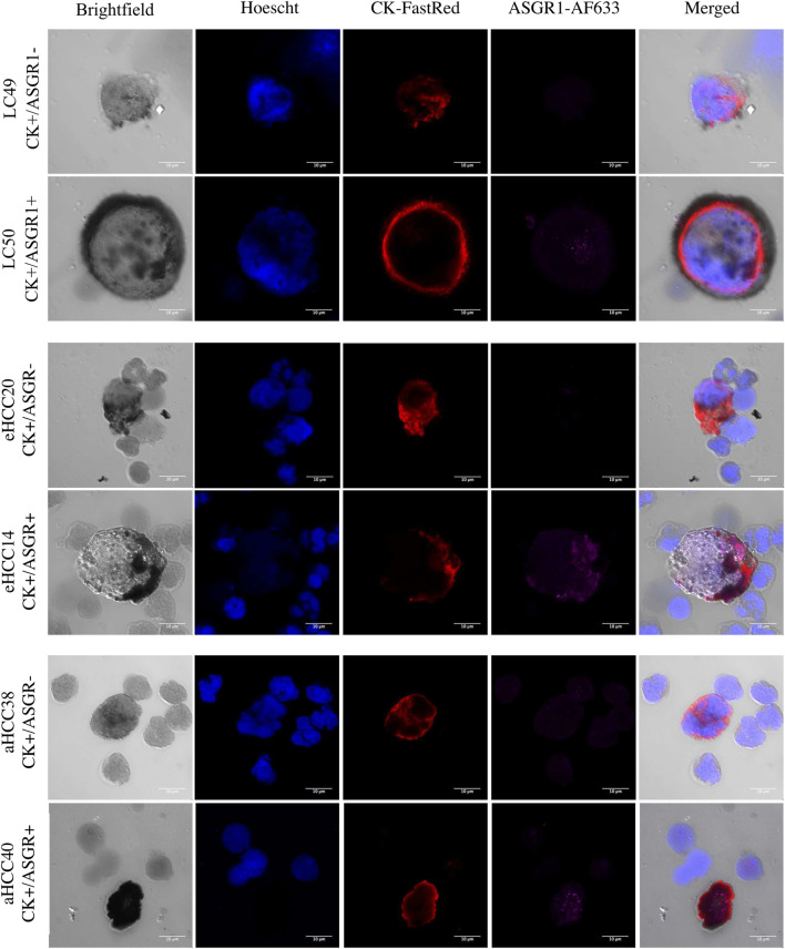
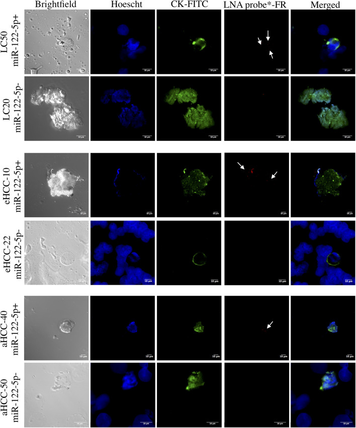
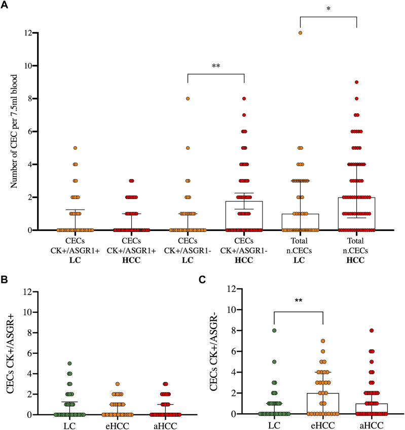
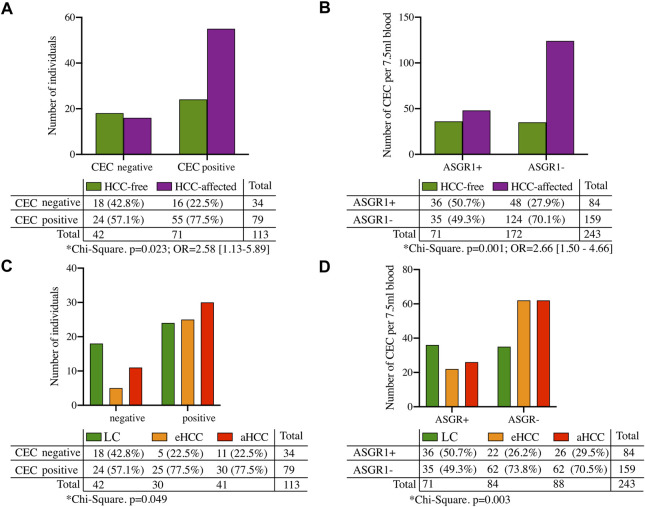
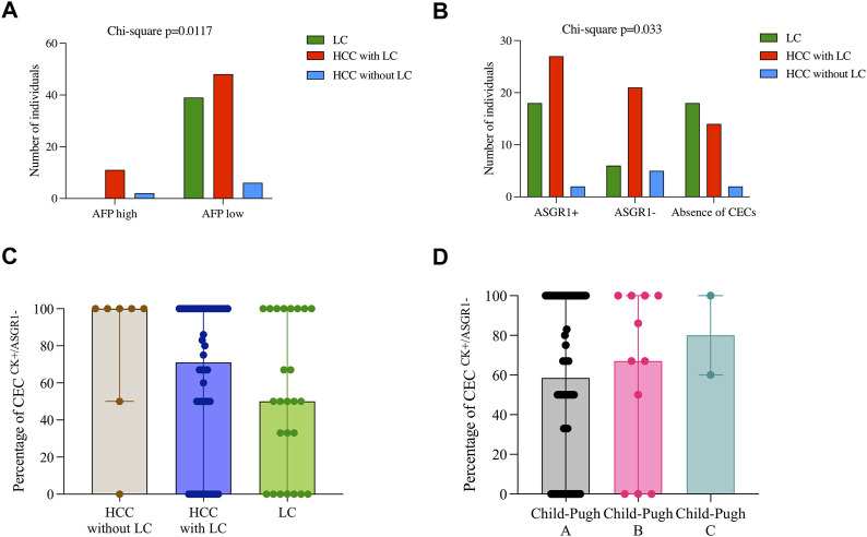
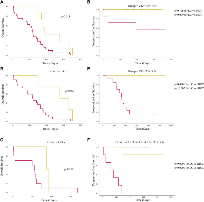
Purpose: Lack of diagnostic and prognostic biomarkers in hepatocellular carcinoma impedes stratifying patients based on their risk of developing cancer. The aim of this study was to evaluate phenotypic and genetic heterogeneity of circulating epithelial cells (CECs) based on asialoglycoprotein receptor 1 (ASGR1) and miR-122-5p expression as potential diagnostic and prognostic tools in patients with hepatocellular carcinoma (HCC) and liver cirrhosis (LC). Methods: Peripheral blood samples were extracted from LC and HCC patients at different disease stages. CECs were isolated using positive immunomagnetic selection. Genetic and phenotypic characterization was validated by double immunocytochemistry for cytokeratin (CK) and ASGR1 or by in situ hybridization with miR-122-5p and CECs were visualized by confocal microscopy. Results: The presence of CECs increased HCC risk by 2.58-fold, however, this was only significant for patients with previous LC (p = 0.028) and not for those without prior LC (p = 0.23). Furthermore, the number of CECs lacking ASGR1 expression correlated significantly with HCC incidence and absence of miR-122-5p expression (p = 0.014; r = 0.23). Finally, overall survival was significantly greater for patients at earlier cancer stages (p = 0.018), but this difference was only maintained in the group with the presence of CECs (p = 0.021) whereas progression-free survival was influenced by the absence of ASGR1 expression. Conclusion: Identification and characterization of CECs by ASGR1 and/or miR-122-5p expression may be used as a risk-stratification tool in LC patients, as it was shown to be an independent prognostic and risk-stratification marker in LC and early disease stage HCC patients.

Keywords: cancer interception; circulating tumor cells; hepatocellular carcinoma; liver cirrhosis; precision medicine.


Conflict of interest statement

The authors declare that the research was conducted in the absence of any commercial or financial relationships that could be construed as a potential conflict of interest.

Figures

FIGURE 1
FIGURE 1
CECs heterogeneity in patients with liver cirrhosis or HCC. Heterogeneity is shown with respect to Cytokeratin (CK) and ASGR1 expression in two LC (liver cirrhosis) patients (top), two eHCC (early HCC) patients (middle) and two aHCC (advanced HCC) patients (bottom). In each case, one CEC positive for the two markers (CK and ASGR1) and one CEC negative for ASGR1 are shown. Hoechst is used as nuclear staining.
FIGURE 2
FIGURE 2
Confocal microscopy of immunoFISH for miR-122-5p in patients with liver cirrhosis or HCC. Cytokeratin (CK) is shown in green (FITC) and miR122-5p probe is shown in red. Data from two patients for each group: liver cirrhosis (LC; top), early HCC (eHCC; middle) and advanced HCC (aHCC; bottom) are shown. For each patient group, a positive miR-122-5p signal (coinciding with positive ASGR1 staining) and a negative miR-122-5p signal (coinciding with negative ASGR1 staining) are shown.
FIGURE 3
FIGURE 3
CECs enumeration in blood in patients with liver cirrhosis or HCC. CECs are characterized by either double staining (CK+/ASGR1+) or single staining (CK+/ASGR1-) in liver cirrhosis (LC) and hepatocellular carcinoma (HCC) patients (A). The latter are divided into early (eHCC) and advanced (aHCC) disease for both, double (B) and single staining (C). p values for Kruskal Wallis test and multiple comparisons are:**p = 0.0096 and *p = 0.035 (A) and **p = 0.0051 (C).
FIGURE 4
FIGURE 4
Risk factors for HCC. (A) Shows presence/absence of CECs and (B) shows number of CECs expressing (or not) ASGR1 in liver cirrhosis (LC) or hepatocellular carcinoma patients (HCC). (C,D) show same information as in (A,B) respectively, but dividing HCC individuals by either early (eHCC) or advanced (aHCC) disease stages.
FIGURE 5
FIGURE 5
Relationship between CEC characterization and clinical and pathological features in patients with liver cirrhosis or HCC. (A) Shows AFP levels divided by high (>400 ng/ml) and low (<400 ng/ml) for three groups of patients: LC = liver cirrhosis; HCC of cirrhosis etiology and HCC of other a etiology. (B) Shows frequency distribution of CECs for the same groups of patients. (C,D) Represent percentages of CECs with lack of ASGR1 expression for either the three previous group of patients (C) or Child-Pugh scores (D). p values for Chi-Square tests are shown for graphs (A,B). No significant differences are shown in C and D for Kruskal Wallis tests.
FIGURE 6
FIGURE 6
Survival curves for patients with liver cirrhosis or HCC accounting for CEC numbers and phenotype. Left part of the figure represents overall survival curves for the whole population (A), only for those positive for CECs (B) and for those negative for CECs (C). Colors are Grey = eHCC and Black: aHCC. Right part of the figure represents progression-free survival for patients with all CECs positive for ASGR1 (D), patients with all CECs negative for ASGR1 (E) and patients with heterogeneous CEC phenotypes (F). Colors are: Dotted Grey = liver cirrhosis; Grey eHCC and Black: aHCC.

References

    1. Bayarri-Lara C., Ortega F. G., Cueto Ladrón de Guevara A., Puche J. L., Ruiz Zafra J., de Miguel-Perez D., et al. (2016). Circulating tumor cells identify early recurrence in patients with non-small cell lung cancer undergoing radical resection. PLoS One 11, e0148659. 10.1371/journal.pone.0148659 - DOI - PMC - PubMed
    1. Bialecki E. S., di Bisceglie A. M. (2005). Diagnosis of hepatocellular carcinoma. HPB 7, 26–34. 10.1080/13651820410024049 - DOI - PMC - PubMed
    1. Cadier B., Bulsei J., Nahon P., Seror O., Laurent A., Rosa I., et al. (2017). Early detection and curative treatment of hepatocellular carcinoma: A cost-effectiveness analysis in France and in the United States. Hepatology 65, 1237–1248. 10.1002/hep.28961 - DOI - PubMed
    1. Cancer Tomorrow (2022). Cancer Tomorrow. Available at: https://gco.iarc.fr/tomorrow/en/dataviz/isotype?cancers=11&single_unit=5... . [Accessed 22 Sep 2022].
    1. Carr B. I., Akkiz H., Üsküdar O., Yalcin K., Guerra V., Kuran S., et al. (2018). HCC with low- and normal-serum alpha-fetoprotein levels. Clin. Pract. 15, 453–464. 10.4172/clinical-practice.1000393 - DOI - PMC - PubMed