

Tyrosine bioconjugation with hypervalent iodine
- PMID: 36519034
- PMCID: PMC9645396
- DOI: 10.1039/d2sc04558c
Tyrosine bioconjugation with hypervalent iodine
Erratum in
-
Correction: Tyrosine bioconjugation with hypervalent iodine.Chem Sci. 2022 Dec 15;14(2):393-394. doi: 10.1039/d2sc90252d. eCollection 2023 Jan 4. Chem Sci. 2022. PMID: 36687350 Free PMC article.
Abstract
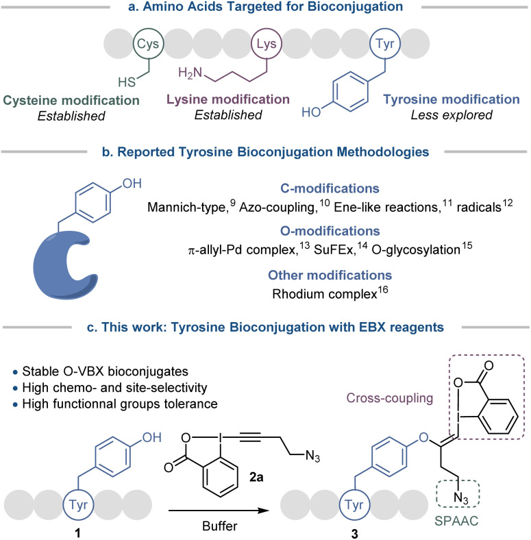
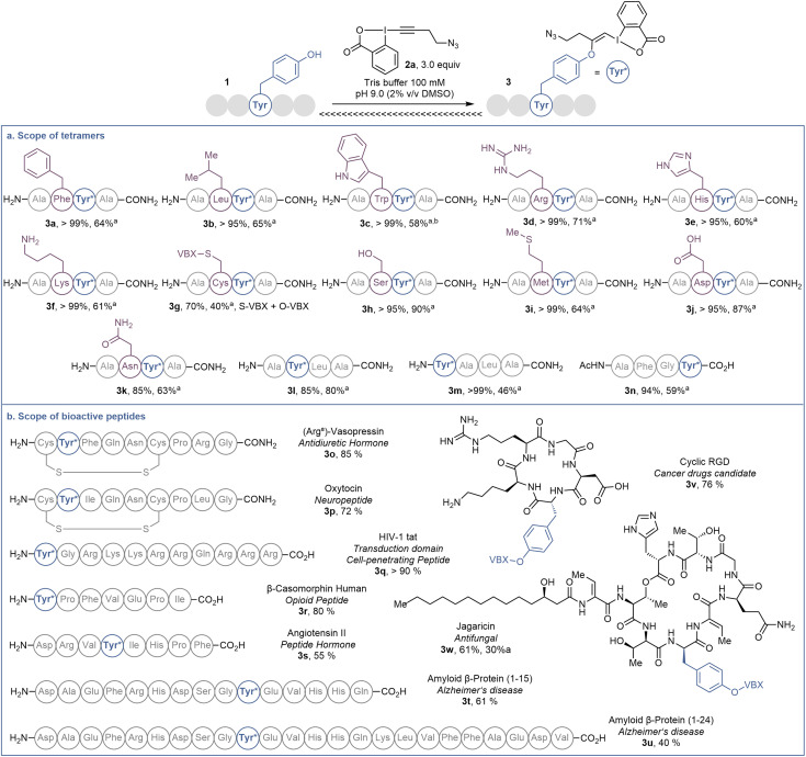
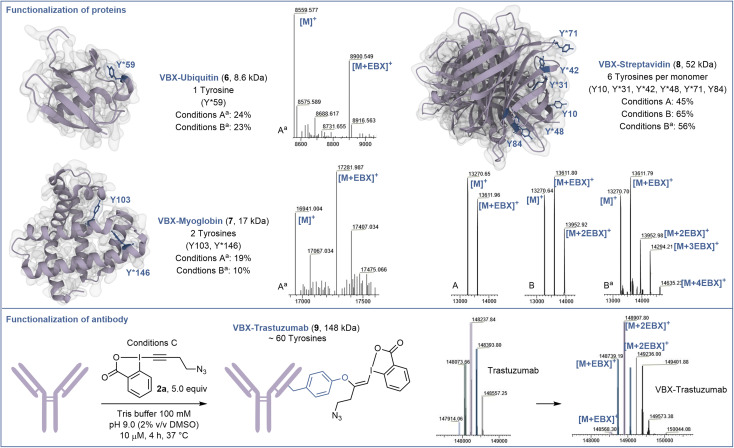
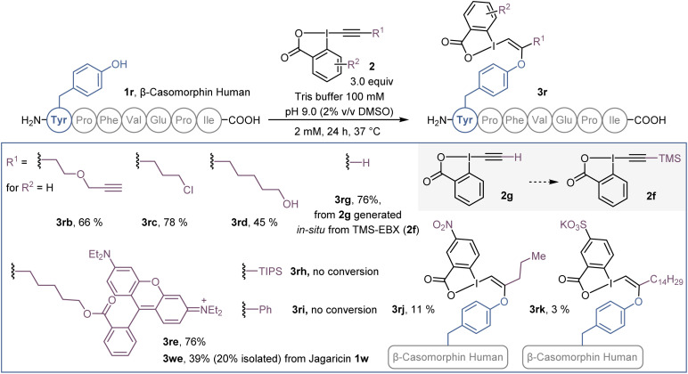
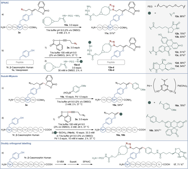
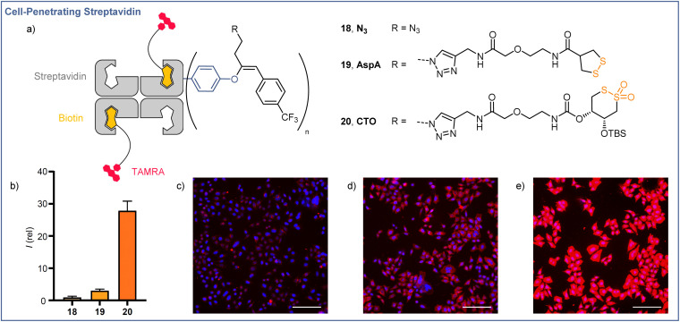
Hypervalent iodine reagents have recently emerged as powerful tools for late-stage peptide and protein functionalization. Herein we report a tyrosine bioconjugation methodology for the introduction of hypervalent iodine onto biomolecules under physiological conditions. Tyrosine residues were engaged in a selective addition onto the alkynyl bond of ethynylbenziodoxolones (EBX), resulting in stable vinylbenziodoxolones (VBX) bioconjugates. The methodology was successfully applied to peptides and proteins and tolerated all other nucleophilic residues, with the exception of cysteine. The generated VBX were further functionalized by palladium-catalyzed cross-coupling and azide-alkyne cycloaddition reactions. The method could be successfully used to modify bioactive natural products and native streptavidin to enable thiol-mediated cellular uptake.
This journal is © The Royal Society of Chemistry.
Conflict of interest statement
There are no conflicts to declare.
Figures
References
-
- Fosgerau K. Hoffmann T. Drug Discovery Today. 2015;20:122. doi: 10.1016/j.drudis.2014.10.003. - DOI - PubMed
- Hu Q.-Y. Berti F. Adamo R. Chem. Soc. Rev. 2016;45:1691–1719. doi: 10.1039/C4CS00388H. - DOI - PubMed
- Gunnoo S. B. Madder A. Org. Biomol. Chem. 2016;14:8002–8013. doi: 10.1039/C6OB00808A. - DOI - PubMed
- Lau J. L. Dunn M. K. Bioorg. Med. Chem. 2018;26:2700–2707. doi: 10.1016/j.bmc.2017.06.052. - DOI - PubMed
-
- Spicer C. D. Davis B. G. Nat. Commun. 2014;5:4740. doi: 10.1038/ncomms5740. - DOI - PubMed
- Koniev O. Wagner A. Chem. Soc. Rev. 2015;44:5495–5551. doi: 10.1039/C5CS00048C. - DOI - PubMed
- Boutureira O. Bernardes G. J. L. Chem. Rev. 2015;115:2174–2195. doi: 10.1021/cr500399p. - DOI - PubMed
- Hoyt E. A. Cal P. M. S. D. Oliveira B. L. Bernardes G. J. L. Nat. Rev. Chem. 2019;3:147–171. doi: 10.1038/s41570-019-0079-1. - DOI
- Tamura T. Hamachi I. J. Am. Chem. Soc. 2019;141:2782–2799. doi: 10.1021/jacs.8b11747. - DOI - PubMed
- Boto A. González C. C. Hernández D. Romero-Estudillo I. Saavedra C. J. Org. Chem. Front. 2021;8:6720–6759. doi: 10.1039/D1QO00892G. - DOI
- Walsh S. J. Bargh J. D. Dannheim F. M. Hanby A. R. Seki H. Counsell A. J. Ou X. Fowler E. Ashman N. Takada Y. et al. Chem. Soc. Rev. 2021;50:1305–1353. doi: 10.1039/D0CS00310G. - DOI - PubMed
- Sornay C. Vaur V. Wagner A. Chaubet G. R. Soc. Open Sci. 2022;9:211563. doi: 10.1098/rsos.211563. - DOI - PMC - PubMed
-
- Lin S. Yang X. Jia S. Weeks A. M. Hornsby M. Lee P. S. Nichiporuk R. V. Iavarone A. T. Wells J. A. Toste F. D. et al. Science. 2017;355:597–602. doi: 10.1126/science.aal3316. - DOI - PMC - PubMed
- Taylor M. T. Nelson J. E. Suero M. G. Gaunt M. J. Nature. 2018;562:563–568. doi: 10.1038/s41586-018-0608-y. - DOI - PMC - PubMed
- Kim J. Li B. X. Huang R. Y.-C. Qiao J. X. Ewing W. R. MacMillan D. W. C. J. Am. Chem. Soc. 2020;142:21260–21266. doi: 10.1021/jacs.0c09926. - DOI - PMC - PubMed
-
- Antos J. M. Francis M. B. J. Am. Chem. Soc. 2004;126:10256–10257. doi: 10.1021/ja047272c. - DOI - PubMed
- Seki Y. Ishiyama T. Sasaki D. Abe J. Sohma Y. Oisaki K. Kanai M. J. Am. Chem. Soc. 2016;138:10798–10801. doi: 10.1021/jacs.6b06692. - DOI - PubMed
- Tower S. J. Hetcher W. J. Myers T. E. Kuehl N. J. Taylor M. T. J. Am. Chem. Soc. 2020;142:9112–9118. doi: 10.1021/jacs.0c03039. - DOI - PMC - PubMed
LinkOut - more resources
Full Text Sources