. 2023 Apr 25;33(9):5613-5624.
doi: 10.1093/cercor/bhac446.

Fetal development of functional thalamocortical and cortico-cortical connectivity

Affiliations

Fetal development of functional thalamocortical and cortico-cortical connectivity

Athena Taymourtash et al. Cereb Cortex. .

Abstract

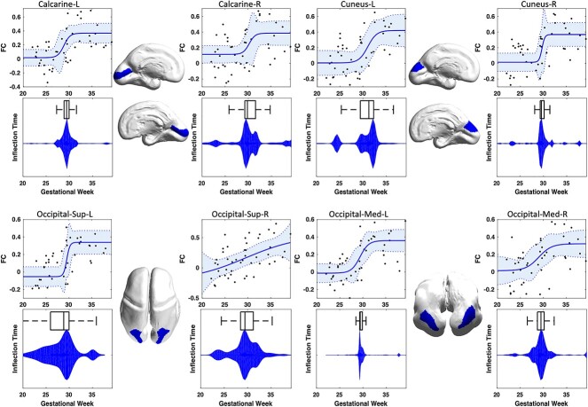
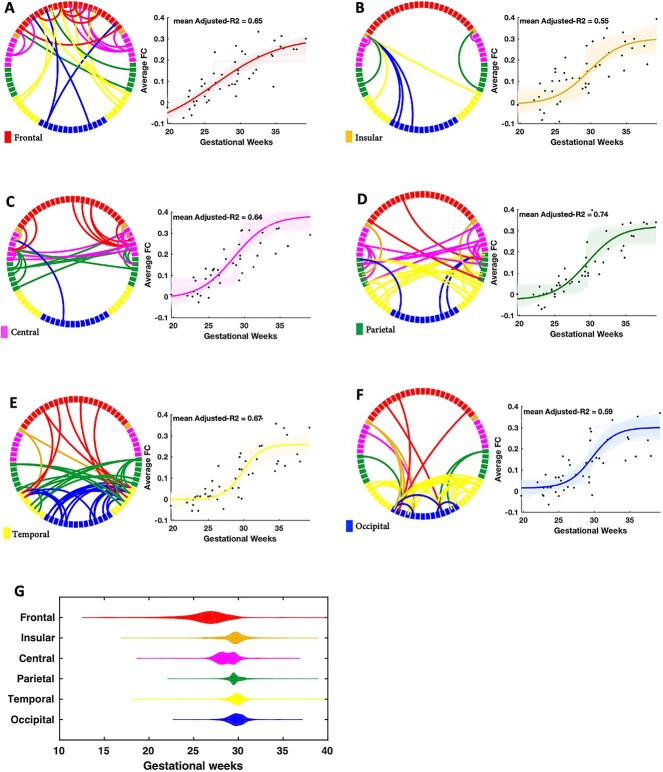
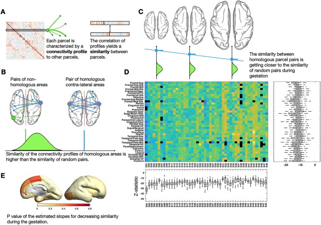
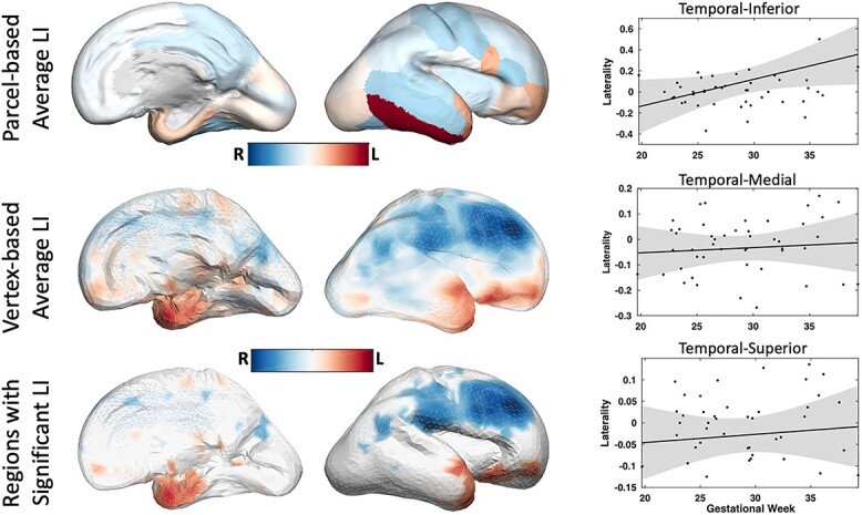
Measuring and understanding functional fetal brain development in utero is critical for the study of the developmental foundations of our cognitive abilities, possible early detection of disorders, and their prevention. Thalamocortical connections are an intricate component of shaping the cortical layout, but so far, only ex-vivo studies provide evidence of how axons enter the sub-plate and cortex during this highly dynamic phase. Evidence for normal in-utero development of the functional thalamocortical connectome in humans is missing. Here, we modeled fetal functional thalamocortical connectome development using in-utero functional magnetic resonance imaging in fetuses observed from 19th to 40th weeks of gestation (GW). We observed a peak increase of thalamocortical functional connectivity strength between 29th and 31st GW, right before axons establish synapses in the cortex. The cortico-cortical connectivity increases in a similar time window, and exhibits significant functional laterality in temporal-superior, -medial, and -inferior areas. Homologous regions exhibit overall similar mirrored connectivity profiles, but this similarity decreases during gestation giving way to a more diverse cortical interconnectedness. Our results complement the understanding of structural development of the human connectome and may serve as the basis for the investigation of disease and deviations from a normal developmental trajectory of connectivity development.

Keywords: fetal brain development; functional connectivity; magnetic resonance imaging; thalamus.


Figures

Fig. 1
Fig. 1
Single subject processing pipeline from raw fMRI data to subject-specific functional connectivity map. A) correction of signal nonuniformity; B) motion correction; C) rejection of outlier volumes, nuisance regression, and temporal filtering; D) super-resolution 3D reconstruction of the raw T2 images and the resulting segmentation; E) registration of the segmentation to the fMRI space, clean bold signal extracted from each segment (by averaging across voxels), resulting functional connectivity map, and illustration of the cortical lobes on a customized fetal surface.
Fig. 2
Fig. 2
Association between the strength of thalamocortical FC and gestational age of fetuses. Each panel shows the estimated nonlinear growth trajectory, the distribution of the achieved inflection times, and the projection of each region onto a surface rendering of a 26-weeks-old fetal brain.
Fig. 3
Fig. 3
Development of cortico–cortical FC across gestational age of fetuses. Each circular graph depicts one cortical lobe with restricted edges whose development showed a significant correlation with gestational age after correcting for multiple comparisons. The violin plots show the distribution of the estimated inflection times for each cortical lobe.
Fig. 4
Fig. 4
Similarity analysis. For a single subject, a similarity map was was quantified as Pearson’s correlation coefficient between their functional connectivity profiles (A) and was tested for homologous parcels (B). We observed the similarity between homologous parcels is significantly higher than the similarity between random pairs of parcels; however, it decreases during the gestation (C). This analysis was repeated for all parcels of all fetuses and the test-statistics (sorted by age) is shown in (D). P-values of the estimated slopes for decreasing similarity during gestation is demonstrated in (E).
Fig. 5
Fig. 5
Multi-subject maps of brain functional asymmetry. Image-analysis techniques make it possible to distinguish systematic functional asymmetries in a population, or a specific group of subjects, from random fluctuations in functional connectivity. Average LI values across 48 normal fetuses are shown in color which reveals prominent asymmetries in Temporal-Inferior, Temporal-Medial, and Temporal-Superior areas. By comparing the average magnitude of these asymmetries with their standard error—derived from the standard deviation—regions of significant asymmetry are identified.

References

    1. Alcauter S, Weili Lin J, Smith K, Short SJ, Goldman BD, Steven Reznick J, Gilmore JH, Gao W. Development of thalamocortical connectivity during infancy and its cognitive correlations. J Neurosci. 2014:34(27):9067–9075. - PMC - PubMed
    1. Balice-Gordon RJ, Lichtman JW. Long-term synapse loss induced by focal blockade of postsynaptlc receptors. Nature. 1994:372(6506):519–524. - PubMed
    1. Ball G, Boardman JP, Aljabar P, Pandit A, Arichi T, Merchant N, Daniel Rueckert A, Edwards D, Counsell SJ. The influence of preterm birth on the developing thalamocortical connectome. Cortex. 2013:49(6):1711–1721. - PubMed
    1. Ball G, Pazderova L, Chew A, Tusor N, Merchant N, Arichi T, Allsop JM, Cowan FM, David Edwards A, Counsell SJ. Thalamocortical connectivity predicts cognition in children born preterm. Cereb Cortex. 2015:25(11):4310–4318. - PMC - PubMed
    1. Behzadi Y, Restom K, Liau J, Liu TT. A component based noise correction method (compcor) for bold and perfusion based fmri. NeuroImage. 2007:37(1):90–101. - PMC - PubMed

Publication types