. 2022 Dec 16;378(6625):1227-1234.
doi: 10.1126/science.ade0156. Epub 2022 Dec 15.

Multidimensional control of therapeutic human cell function with synthetic gene circuits

Affiliations

Multidimensional control of therapeutic human cell function with synthetic gene circuits

Hui-Shan Li et al. Science. .

Abstract

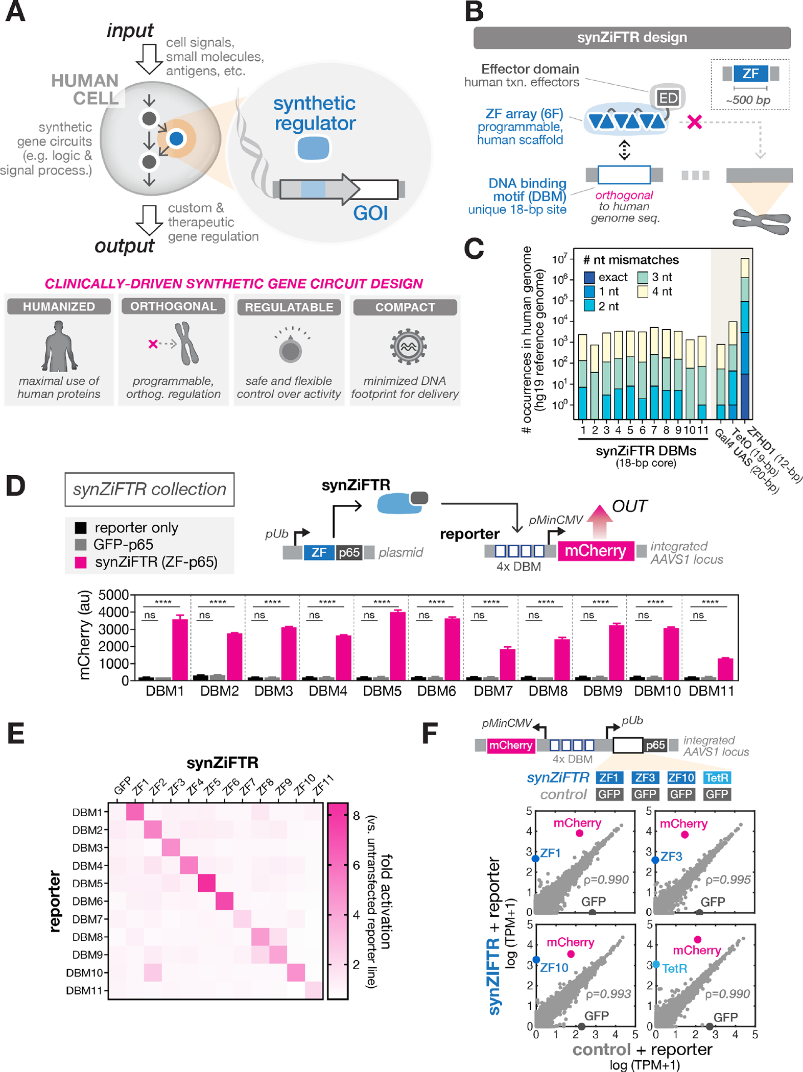
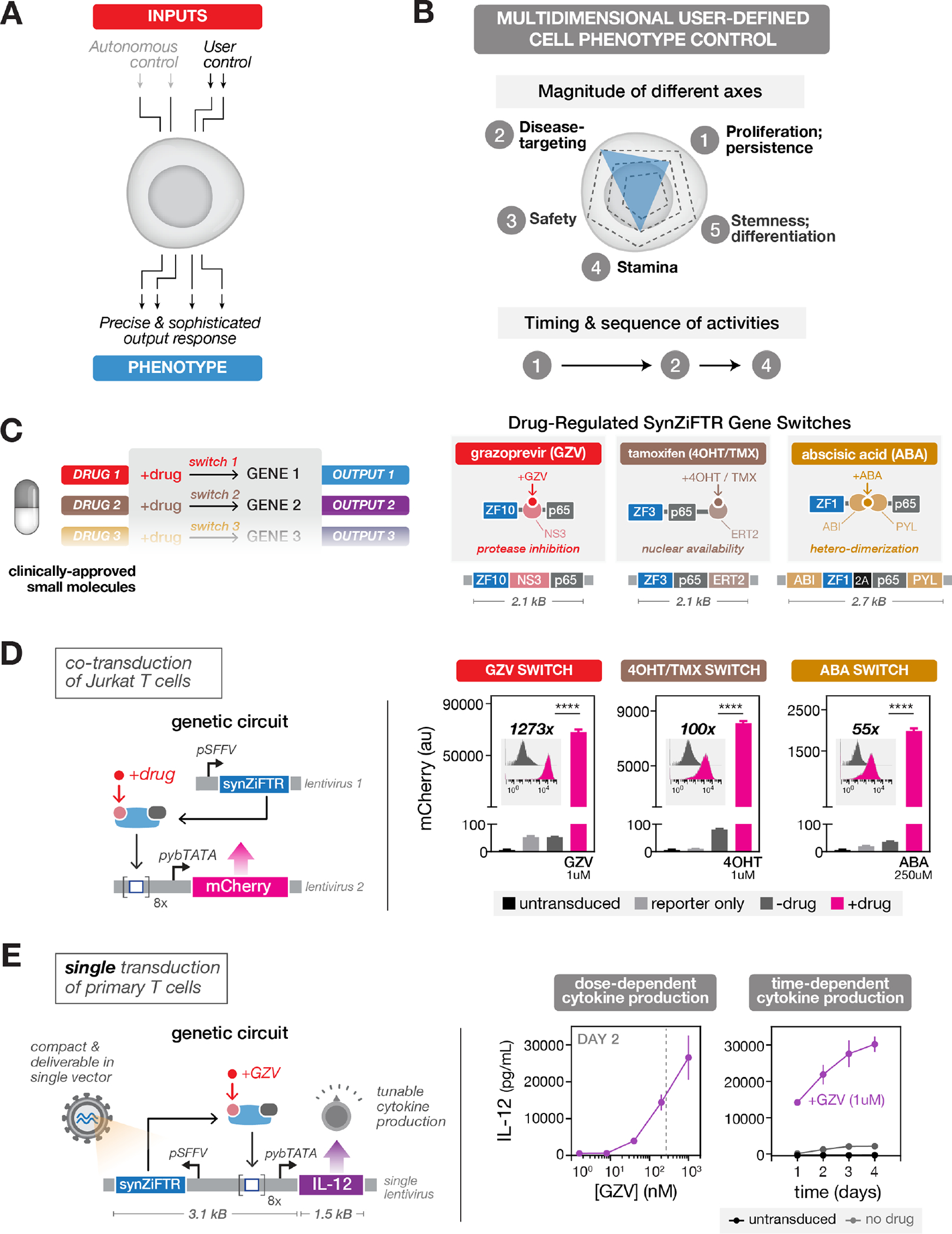
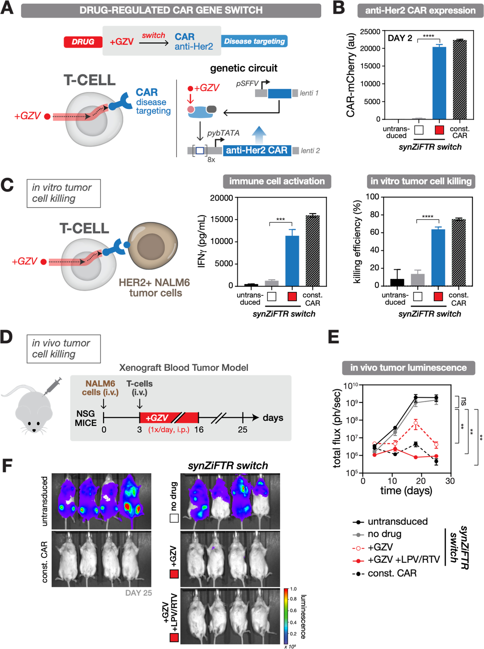
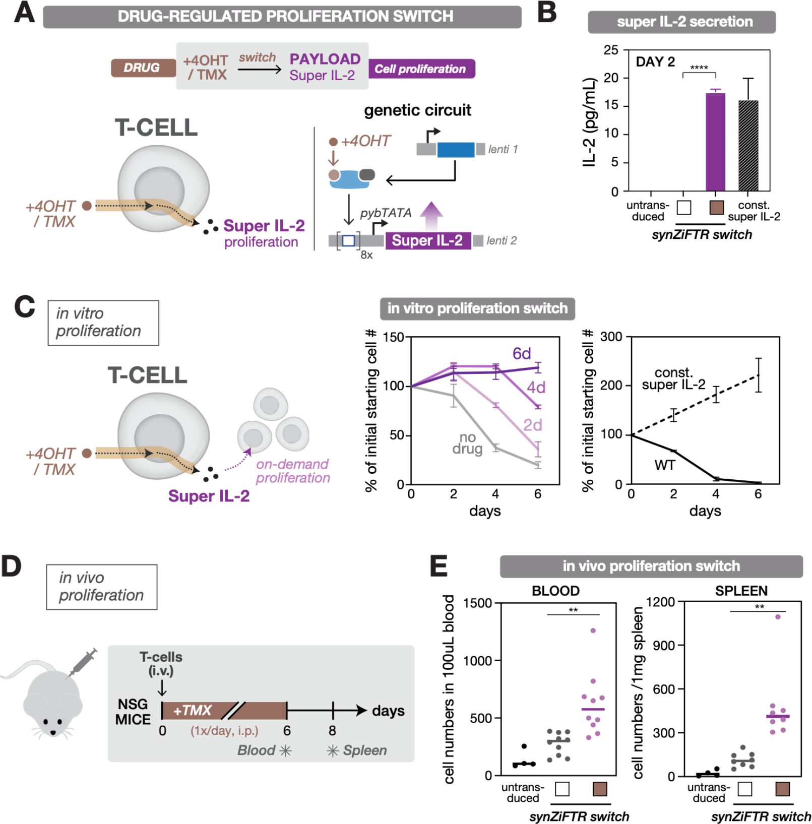
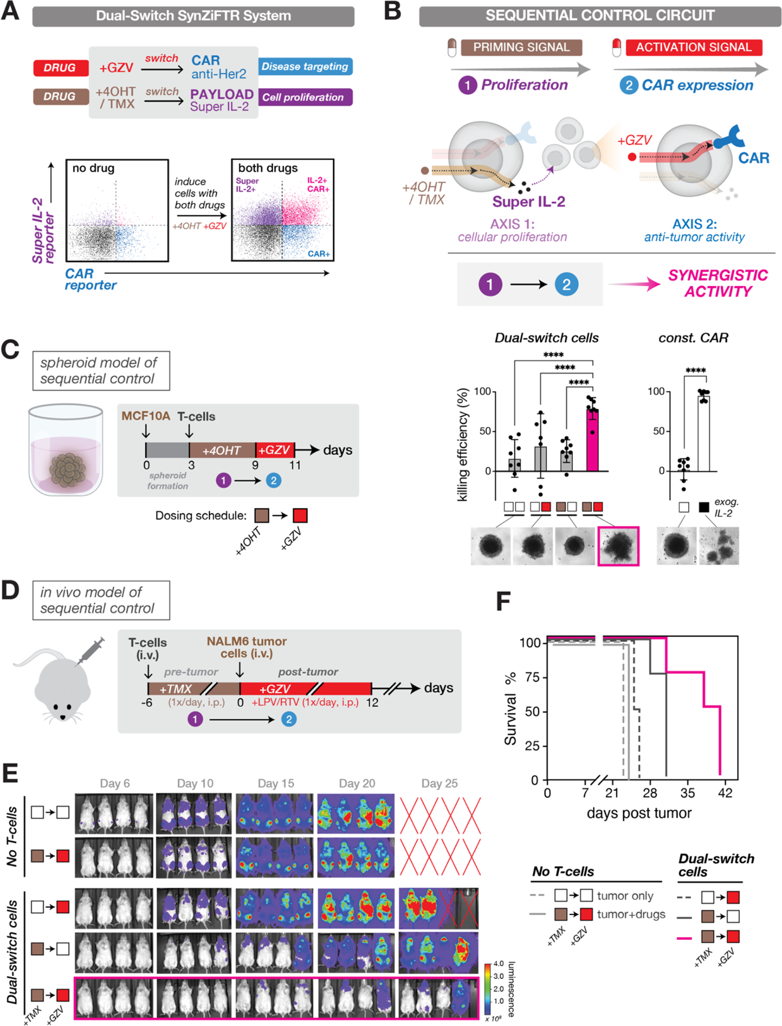
Synthetic gene circuits that precisely control human cell function could expand the capabilities of gene- and cell-based therapies. However, platforms for developing circuits in primary human cells that drive robust functional changes in vivo and have compositions suitable for clinical use are lacking. Here, we developed synthetic zinc finger transcription regulators (synZiFTRs), which are compact and based largely on human-derived proteins. As a proof of principle, we engineered gene switches and circuits that allow precise, user-defined control over therapeutically relevant genes in primary T cells using orthogonal, US Food and Drug Administration-approved small-molecule inducers. Our circuits can instruct T cells to sequentially activate multiple cellular programs such as proliferation and antitumor activity to drive synergistic therapeutic responses. This platform should accelerate the development and clinical translation of synthetic gene circuits in diverse human cell types and contexts.


Conflict of interest statement

Competing interests: D.V.I., J.D.S., J.K.J, and A.S.K. are inventors on a patent related to the synZiFTR technology; H.S.L., D.V.I., K.T.R., W.W.W, and A.S.K. have filed patent applications related to drug-regulated synZiFTRs. J.K.J. is a co-inventor on various patents and patent applications that describe gene editing and epigenetic editing technologies. K.T.R. is a co-founder of Arsenal Biosciences, was a founding scientist/consultant and stockholder in Cell Design Labs, now a Gilead Company, and holds stock in Gilead. J.K.J. has, or had during the course of this research, financial interests in several companies developing gene editing technology: Beam Therapeutics, Blink Therapeutics, Chroma Medicine, Editas Medicine, EpiLogic Therapeutics, Excelsior Genomics, Hera Biolabs, Monitor Biotechnologies, Nvelop Therapeutics (f/k/a ETx, Inc.), Pairwise Plants, Poseida Therapeutics, SeQure Dx, Inc., and Verve Therapeutics. J.K.J.’s financial interests in these companies include consulting fees and/or equity. J.K.J.’s interests were reviewed and are managed by Massachusetts General Hospital and Mass General Brigham in accordance with their conflict of interest policies. W.W.W. is a scientific co-founder of and holds equity in Senti Biosciences. A.S.K. is a scientific advisor for and holds equity in Senti Biosciences and Chroma Medicine, and is a co-founder of Fynch Biosciences and K2 Biotechnologies.

Figures

Fig. 1.
Fig. 1.. Clinically-driven design of compact, humanized, synthetic gene regulators (synZiFTRs) for mammalian cell engineering
(A) Synthetic gene circuits are used to convert diverse input signals into desired gene expression outputs in order to precisely control human cell function (top). Criteria for clinically-driven gene circuit design framework (bottom). (B) SynZiFTR design. SynZiFTRs have a modular design based on compact, human-derived protein domains. An engineered ZF array mediates interactions with a unique, human genome-orthogonal DNA-binding motif (DBM), and human-derived effector domains (EDs) are used to modulate transcriptional activity. (C) Prevalence of synZiFTR recognition motifs in the human genome. Occurrences of exact and increasingly mismatched sequences for each synZiFTR DBM and response elements from common artificial regulators (Gal4 UAS, TetO, ZFHD1). (D) SynZiFTRs strongly activate gene expression at corresponding response promoters. Response element vectors were stably-integrated into HEK293FT cells to generate reporter lines for each synZiFTR (ZF-p65 fusion). SynZiFTR (or control) expression vectors were transfected into corresponding reporter lines, and mCherry was measured by flow cytometry after 2 days. Bars represent mean values for three measurements ± SD. Statistics represent one-way ANOVA with Dunnett’s Multiple Comparisons; ns: not significant; ****: p < 0.0001. pUb, Ubiquitin C promoter; pMinCMV, minimal CMV promoter; p65, aa361-551. (E) SynZiFTRs have mutually orthogonal regulatory specificities. Each synZiFTR expression vector was transfected into every reporter line, and mCherry was measured by flow cytometry after 2 days. Fold activation levels represent mean values for three biological replicates. (F) SynZiFTRs exhibit specific and orthogonal transcriptional regulation profiles in human cells. Correlation of transcriptomes from RNA-sequencing measurements of HEK293FT cells stably expressing synZiFTR or TetR-p65 versus a GFP-p65 control. Points represent individual transcript levels normalized to TPM, transcripts per kilobase million, averaged between two technical replicates. Pearson correlation coefficient was calculated for native (grey) transcripts. See Fig. S3 for extended analyses.
Fig. 2.
Fig. 2.. SynZiFTR gene switches allow precise, user-defined control over gene expression in human cells using clinically-approved small molecules
(A) Two forms of cellular control: circuits can be designed to enact cell-autonomous phenotype control (e.g., via recognition of disease-relevant cell surface molecules) or external, user-defined phenotype control (e.g., via administration of small molecules). (B) User-defined control over different axes of a cellular phenotype (top) and the chronology of cellular activities (bottom). (C) Implementing multi-gene user control with orthogonal gene switches that are regulated by clinically-viable small molecules (right). Design of three distinct synZiFTR gene switches that are controlled by orthogonal small molecules: grazoprevir (GZV), 4-hydroxytamoxifen / tamoxifen (4OHT / TMX), and abscisic acid (ABA) (right). NS3, hepatitis C virus NS3 protease domain; ERT2, human estrogen receptor T2 mutant domain; ABI, ABA-insensitive 1 domain (aa 126–423); PYL, PYR1-like 1 domain (aa 33–209). (D) Optimized synZiFTR switches enable strong inducible gene expression in Jurkat T cells. Jurkat T cells were co-transduced with reporter and synZiFTR expression lentiviral vectors in an equal ratio. mCherry fluorescence was measured by flow cytometry 4 days following induction by small molecules at indicated concentrations. Bars represent mean values for three measurements ± SD. Statistics represent two-tailed Student’s t test; ***: p < 0.001; ****: p < 0.0001. Histograms show absolute levels and mean fold activation for one representative measurement (insets). pSFFV, Spleen Focus-Forming Virus promoter; pybTATA, synthetic YB_TATA promoter. (E) Compact, single lentivirus-encoded synZiFTR switches enable titratable control over the expression of therapeutically-relevant genes in primary human immune cells. Human primary T cells were transduced with a single lentiviral vector encoding GZV-regulated IL-12 (see Methods). IL-12 production was measured by ELISA at specified time points following induction (with or without 1 uM GZV). Points represent mean values for three measurements ± SD. Dashed line, estimate of the Cmax for traditional clinical dosing of GZV.
Fig. 3.
Fig. 3.. SynZiFTR gene circuit for drug-regulated, post-delivery control over CAR expression and T-cell killing in vivo
(A) Design of the synZiFTR gene circuit for GZV-dependent control over anti-Her2 CAR expression and tumor cell targeting and killing. (B) GZV-regulated CAR expression in primary T cells. Human primary T cells were co-transduced with equal ratios of lentiviral vectors encoding the synZiFTR CAR gene circuit (see Methods). Expression of anti-Her2 CAR-mCherry was measured by flow cytometry two days following induction (with or without 1 uM GZV). White box, uninduced; red box, GZV induced. Const. CAR, constitutively expressed (pSFFV-CAR). Bars represent mean values for three measurements ± SD. Statistics represent two-tailed Student’s t test; ***: p < 0.001; ****: p < 0.0001. (C) GZV-regulated immune cell activation and tumor cell killing in vitro. SynZiFTR-controlled CAR T cells (pre-induced with or without 1 uM GZV for 2 days) were co-cultured with HER2+ NALM6 target leukemia cells in a 1:1 ratio (left). IFNγ secretion from activated immune cells was measured by ELISA (center) and tumor cell killing by flow cytometry (right), one day following co-culturing. White box, uninduced; red box, GZV induced. (D) Testing in vivo efficacy of synZiFTR-regulated CAR T cells using a xenograft tumor mouse model. Timeline of in vivo experiment, in which NSG mice were injected i.v. with luciferase-labeled HER2+ NALM6 cells to establish tumor xenografts, followed by treatment with T cells. GZV was formulated alone or in combination with LPV/RTV and administered i.p. daily over 14 days. Mice were imaged weekly on days 4, 11, 18, 25 to monitor tumor growth via luciferase activity. GZV, 25 mg/kg. LPV/RTV, 10 mg/kg. (E) Tumor burden over time, quantified as the total flux (photons/sec) from the luciferase activity of each mouse using IVIS imaging. Points represent mean values ± SEM (n=4 mice per condition). Statistics represent two-tailed, ratio paired Student’s t test; ns: not significant; **: p < 0.01. (F) IVIS imaging of mouse groups treated with (1) untransduced cells, (2) synZiFTR-regulated CAR T cells, (3) synZiFTR-regulated CAR T cells with GZV, (4) synZiFTR-regulated CAR T cells with GZV+LPV/RTV, (4) constitutive CAR cells. (n=4 mice per condition).
Fig. 4.
Fig. 4.. SynZiFTR gene circuit for drug-regulated, on-demand immune cell proliferation
(A) Design of the synZiFTR gene circuit for 4OHT/TMX-dependent control over super IL-2 expression and cell proliferation. (B) 4OHT-regulated super IL-2 production in primary T cells. Human primary T cells were co-transduced with equal ratios of lentiviral vectors encoding the synZiFTR-regulated proliferation gene circuit (see Methods). Secretion of super IL-2 was measured by ELISA (for IL-2) two days following induction (with or without 1 uM 4OHT). White box, uninduced; brown box, 4OHT induced. Const. super IL-2, constitutively expressed (pSFFV-super IL-2). Bars represent mean values for three measurements ± SD. Statistics represent two-tailed Student’s t-test; ***: p < 0.001; ****: p < 0.0001. (C) 4OHT-regulated T cell proliferation in vitro. SynZiFTR-regulated primary T cells were cultured in IL-2-free media, induced with 4OHT (1 uM) for different durations, and live-cell numbers were quantified by flow cytometry at indicated days (center). Untransduced (WT) and constitutively expressing (const. super IL-2) T cells were cultured and quantified similarly (right). Lines represent mean values for three measurements ± SD. (D) Testing in vivo efficacy of the drug-regulated, on-demand proliferation switch. Timeline of in vivo experiment, in which NSG mice were injected i.v. with primary T cells followed by daily treatment with TMX over six days. The blood and spleen were individually sampled on days 6 and 8, respectively, to quantify the change in T cell numbers. TMX, 75mg/kg. (E) TMX-regulated T cell expansion in vivo. Human T cell numbers from blood and spleen samples were quantified by flow cytometry by gating for hCD3+ / mCD45- cells, following staining for human CD3 (hCD3) and mouse CD45 (mCD45). Lines indicate the mean value; dots represent each mouse. Blood sample (n=10), and spleen sample (n=8). White box, uninduced; brown box, TMX induced. Statistics represent two-tailed Student’s t-test; **: p < 0.01.
Fig. 5.
Fig. 5.. Enacting sequential control of immune cell function to drive synergistic in vivo responses
(A) A dual-switch synZiFTR system for orthogonal, drug-inducible control over anti-Her2 CAR and super IL-2 expression (top). Assessing circuit activation in primary T cells following induction with both drugs (1uM GZV and 1uM 4OHT) (bottom). Distinguishable reporters were used to measure gene activation for each channel with or without 1-day of induction: mCherry-fused anti-Her2 CAR and bicistronic super IL-2 reporter (super IL-2-2A-EGFP). See also Fig. S10. (B) Schema for sequential control in which a priming signal (4OHT/TMX drug) is used to induce cellular proliferation of a small starting population of dual-switch cells via super IL-2 expression, followed by an activation signal (GZV drug) to induce cytotoxic activation via CAR expression. (C) An in vitro spheroid model used to demonstrate the synergistic efficacy of sequential control over T cell proliferation (+4OHT) and activation (+GZV) behavior (left). T cell killing efficiency was measured by luminescence signals from spheroid, and representative morphology of spheroids at the endpoint is shown below (right). White box, uninduced; brown box, 4OHT induced; red box, GZV induced. Bars represent mean values ± SD. Const. CAR, constitutive CAR cells. (D) An in vivo model used to demonstrate the synergistic efficacy of sequential control over T cell proliferation (+TMX) and activation (+GZV) behavior. Timeline of in vivo experiment in which NSG mice were injected i.v. with dual-switch T cells (1×10⁶ cells) six days ahead of the tumor challenge. TMX was administered i.p. daily over six days to activate the proliferation switch prior to tumor challenge. HER2+/Luciferase+ NALM6 cells (1×10⁶ cells) were injected i.v. six days after injection of T cells and the GZV-regulated CAR was switched ON by administering GZV in combination with LPV/RTV daily i.p. over 12 days. TMX, 75mg/kg. GZV, 25mg/kg. LPV/RTV, 10mg/kg. (E) IVIS imaging of tumor burden over time of mouse groups treated with (1) tumor alone (no T cells), (2) tumor with both drugs in sequence (TMX ® GZV), (3) dual-switch T cells treated with GZV alone during the time window of d0 ® d12 (∅ ® GZV), (4) dual-switch T cells treated with TMX alone during the time window of d-6 ® d0 (TMX ® ∅), and (5) dual-switch T cells treated with both drugs in sequence (TMX ® GZV). (F) Kaplan-Meier survival curves for the various treatment groups for the in vivo sequential model study. (n=4 mice per condition). White box, uninduced; brown box, TMX induced; red box, GZV induced.

Comment in

References

    1. Fischbach MA, Bluestone JA, Lim WA, Cell-based therapeutics: the next pillar of medicine. Sci Transl Med 5, 179ps177 (2013). - PMC - PubMed
    1. Kitada T, DiAndreth B, Teague B, Weiss R, Programming gene and engineered-cell therapies with synthetic biology. Science 359, eaad1067 (2018). - PMC - PubMed
    1. Xie M, Fussenegger M, Designing cell function: assembly of synthetic gene circuits for cell biology applications. Nat Rev Mol Cell Biol 19, 507–525 (2018). - PubMed
    1. Lim WA, June CH, The Principles of Engineering Immune Cells to Treat Cancer. Cell 168, 724–740 (2017). - PMC - PubMed
    1. Hay KA, Cytokine release syndrome and neurotoxicity after CD19 chimeric antigen receptor-modified (CAR-) T cell therapy. Br J Haematol 183, 364–374 (2018). - PubMed

Substances