Review
. 2023 Apr 1;103(2):1487-1564.
doi: 10.1152/physrev.00018.2022. Epub 2022 Dec 15.

The enteric nervous system

Affiliations
Review

The enteric nervous system

Keith A Sharkey et al. Physiol Rev. .

Abstract

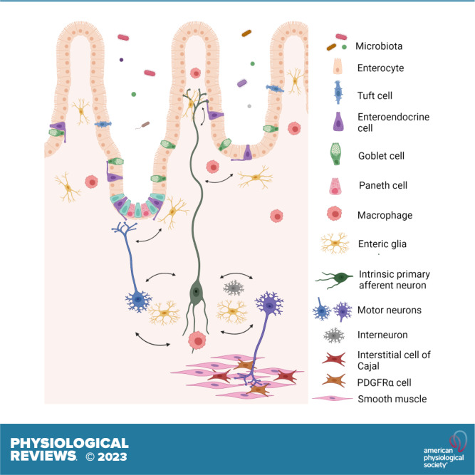
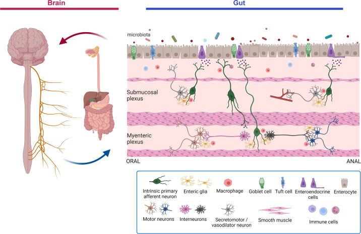
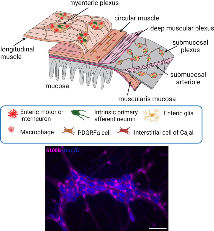
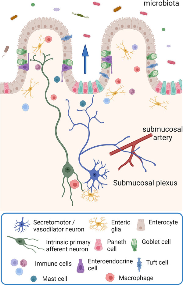
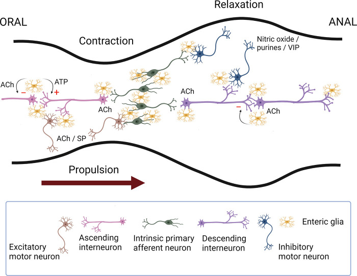
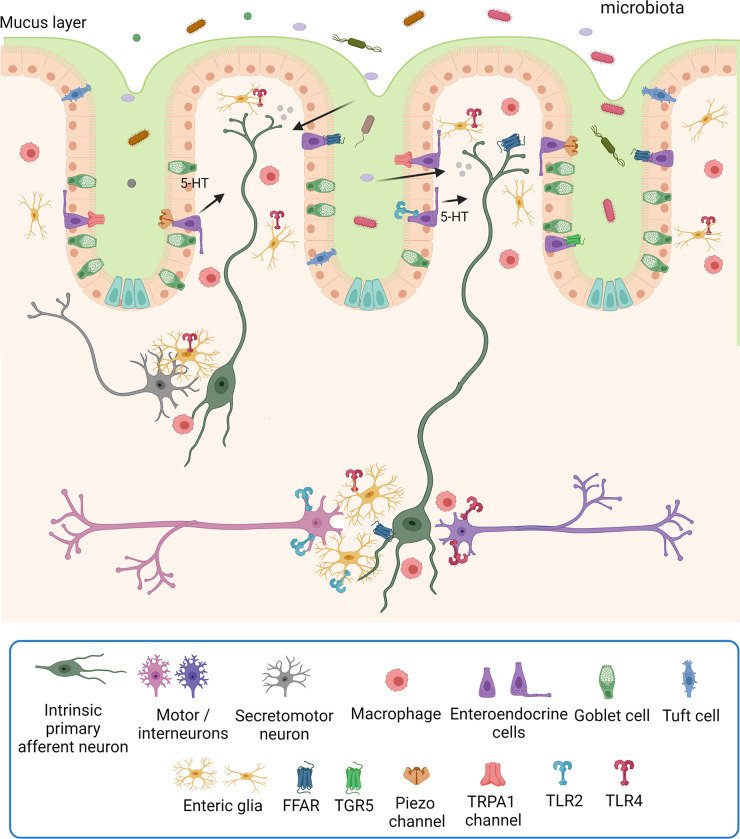
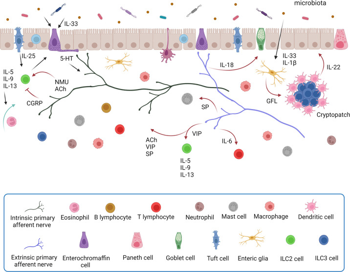
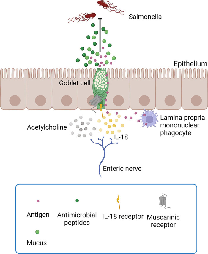
Of all the organ systems in the body, the gastrointestinal tract is the most complicated in terms of the numbers of structures involved, each with different functions, and the numbers and types of signaling molecules utilized. The digestion of food and absorption of nutrients, electrolytes, and water occurs in a hostile luminal environment that contains a large and diverse microbiota. At the core of regulatory control of the digestive and defensive functions of the gastrointestinal tract is the enteric nervous system (ENS), a complex system of neurons and glia in the gut wall. In this review, we discuss 1) the intrinsic neural control of gut functions involved in digestion and 2) how the ENS interacts with the immune system, gut microbiota, and epithelium to maintain mucosal defense and barrier function. We highlight developments that have revolutionized our understanding of the physiology and pathophysiology of enteric neural control. These include a new understanding of the molecular architecture of the ENS, the organization and function of enteric motor circuits, and the roles of enteric glia. We explore the transduction of luminal stimuli by enteroendocrine cells, the regulation of intestinal barrier function by enteric neurons and glia, local immune control by the ENS, and the role of the gut microbiota in regulating the structure and function of the ENS. Multifunctional enteric neurons work together with enteric glial cells, macrophages, interstitial cells, and enteroendocrine cells integrating an array of signals to initiate outputs that are precisely regulated in space and time to control digestion and intestinal homeostasis.

Keywords: enteric glia; enteric nervous system; interstitial cells of Cajal; myenteric plexus; vagus nerve.


Conflict of interest statement

K.A.S. has provided scientific advice and assistance to Arena Pharmaceuticals and GW Pharmaceuticals, has served on a speaker bureau for Abbvie, and has received research support from Takeda Pharmaceuticals and Abalone, Inc. G.M.M. has received research support from Takeda Pharmaceuticals and is on the Scientific Advisory Board of Dignify Therapeutics.

Figures

None
Graphical abstract
Figure 1.
Figure 1.
Schematic overview of the gut-brain-microbiota axis. The gut-brain axis is a bidirectional communication axis that influences the digestive and defensive functions of the gut to regulate homeostasis. The gut microbiota communicates with epithelial cells, including enteroendocrine, tuft cells, and immune cells in the lamina propria that in turn communicate with enteric nerves, enteric glia, and the terminals of extrinsic primary afferent nerves (not shown) to modulate reflex control of gut function. Autonomic reflexes are initiated via vagal and spinal primary afferent pathways, as well as viscerofugal enteric neurons, and central neural circuitry of the brain and spinal cord is mobilized. Top-down control of the gastrointestinal tract occurs via autonomic efferent pathways. Alterations in gut motility and secretion can modulate the microbial environment in the gut. Modifications in the nature of these bidirectional interactions in response to perturbations such as stress or infections can alter the behavior of this system, sometimes manifesting as disorders of brain-gut interactions such as irritable bowel syndrome. Image created with BioRender.com, with permission.
Figure 2.
Figure 2.
Top: the general organization of the enteric nervous system. The myenteric plexus lies between the longitudinal and circular muscle layers. The submucosal plexus lies in the submucosa and in larger animals (not shown) consists of an outer and an inner component. Nerve fibers connect the ganglia and form nerve plexuses that innervate the longitudinal muscle, circular muscle, muscularis mucosa, and mucosa. There are also enteric nerves innervating arteries in the submucosa, as well as the gut-associated lymphoid tissues. The mucosa is densely innervated. Enteric glia are found associated with enteric nerves throughout the wall of the gut. Bottom: organization of enteric neurons (labeled with ant-HuC/D, blue) and enteric glia (labeled with anti-S100B, pink) in the myenteric plexus of the mouse ileum. Note that enteric glia outnumber enteric neurons and the presence of extraganglionic enteric glial cells. Scale bar, 100 µm. See glossary for abbreviations. Unpublished photomicrograph provided by C. M. MacNaughton.
Figure 3.
Figure 3.
The organization of submucosal secretomotor and vasodilator reflexes. Chemical or mechanical stimulation of the mucosa activates enteroendocrine cells, which, in turn, excite submucosal primary afferent neurons that then activate secretomotor and/or vasodilator neurons to stimulate fluid secretion (indicted by the arrow) and/or vasodilatation. Fluid secretion may be amplified by nerve-mediated mast cell activation (13, 135) or modulated by enteric glia (136). Extrinsic primary sensory neurons may also directly evoke a secretory response via axon reflexes (not shown), but there does not appear to be efferent preganglionic input to submucosal ganglia. There are also long secretomotor and vasodilator reflexes involving the myenteric plexus (not shown) (137, 138). Image created with BioRender.com, with permission.
Figure 4.
Figure 4.
Schematic representation of the enteric neural circuitry underlying the neural control of the circular muscle (the main driving force for propulsion). Intrinsic primary afferent neurons synapse with ascending and descending interneurons that run along the length of the intestine, which then activate excitatory (orally) and inhibitory (anally) motor neurons. Upon a chemical or mechanical luminal stimulus, intrinsic primary afferent neurons cause the activation of ascending interneurons that synapse with excitatory motor neurons evoking an oral contraction, whereas the activation of descending interneurons leads to the excitation of inhibitory motor neurons eliciting a relaxation anally. This polarized reflex circuitry allows for a pressure gradient to be developed that propels the contents anally. The primary transmitters involved include acetylcholine (ACh) for all of the pathways except for inhibitory motor neurons that use nitric oxide and purine nucleotides as their primary neurotransmitters. Substance P (SP) or a related tachykinin provides an important adjunct to the excitatory innervation of smooth muscle; likewise, vasoactive intestinal peptide (VIP) is an adjunct to the inhibitory innervation of smooth muscle. Enteric glia also play an active role in regulating intestinal motility. Neuronal recruitment in the descending circuitry is subject to glial inhibitory control in a muscarinic M3-dependent manner. Enteric glial purinergic signaling preferentially stimulates neuronal responses within the ascending circuitry, which may also be under inhibitory regulation (see Ref. for details). Image created with BioRender.com, with permission.
Figure 5.
Figure 5.
Microbiota influence the activity of many cell types in the gut wall, including enteroendocrine cells, tuft cells, immune cells, enteric glia, and enteric neurons, via many different mechanisms including via the production of short-chain fatty acids that activate free fatty acid receptors (FFARs) (235, 236) and bile acids that activate the TGR5 receptor (237), mechanosensitive Piezo1 channels (238), transient receptor potential (TRP)A1 channels (239) and toll-like receptors (TLRs) 2 and 4 (230, 231, 240, 241); see sects. 5.1, 6.2, and 6.5 and FIGURE 9. In addition, the gut microbiota is essential for the maintenance of mucosal enteric glial cells (199), which are key players in mucosal homeostasis (242). Microbial products can directly influence the activity of intrinsic primary afferent neurons (203, 214, 215, 243, 244). See glossary for other abbreviations. Image created with BioRender.com, with permission.
Figure 6.
Figure 6.
A schematic illustration of the interactions between the gut immune and enteric nervous systems in the intestinal mucosa. Bidirectional communication between enteric nerves and immune cells regulates the local environment of the gut. Some of the cellular mediators of neuroimmune signaling in the gut are illustrated. There is extensive multidirectional signaling between intestinal epithelial cells (enterocytes, goblet cells, tuft cells, and enteroendocrine cells), enteric nerves, and enteric glia. Enteric nerves receive and integrate information from enteroendocrine cells, gut microbiota, and enteric glia; they also communicate with enteric glia, epithelia, the microbiota, and various populations of immune cells. Tuft cells and goblet cells are key mediators of epithelial-immune signaling, and tuft cells also signal to enteric nerves. Details of the specific molecular signaling mechanisms are described in the text. A cryptopatch is an aggregate of lymphoid cells found in the intestinal lamina propria. 5-HT, serotonin; ACh, acetylcholine; CGRP, calcitonin gene-related peptide; GFL, glial cell line-derived neurotrophic factor family ligand; IL, interleukin; ILC2, type 2 innate lymphoid cell; ILC3, type 3 innate lymphoid cell; NMU, neuromedin U; SP, substance P; VIP, vasoactive intestinal peptide. Image created with BioRender.com, with permission.
Figure 7.
Figure 7.
Enteric neural regulation of goblet cell secretion is a key element of host defense. Goblet cell secretion of antimicrobial peptides and mucus is differentially regulated by acetylcholine (mediated by mAChR1 muscarinic receptors) and IL-18. Acetylcholine also regulates the passage of antigen through goblet cells by activation of mAChR3 or mAChR4 muscarinic receptors. See text for details. Image created with BioRender.com, with permission.
Figure 8.
Figure 8.
A schematic illustration of the autonomic and primary afferent innervation of the gastrointestinal (GI) tract. The GI tract receives a parasympathetic preganglionic innervation from the vagus and pelvic nerves and a rich sympathetic efferent innervation from postganglionic neurons located in the abdominal prevertebral ganglia (celiac, superior mesenteric, and inferior mesenteric ganglia). Viscerofugal enteric neurons provide input from the small and large intestines to the sympathetic ganglia and from the esophagus to the trachea. Primary afferent neurons carry signals from the gut to the central nervous system. Vagal afferents have their cell bodies in the nodose ganglia, and spinal primary afferent nerves run with the sympathetic and pelvic splanchnic nerves and have their cell bodies in dorsal root ganglia. It should be noted that sympathetic and parasympathetic efferent fibers innervate the enteric nervous system and not the smooth muscle or mucosal tissues of the GI tract, with the exception of a direct sympathetic innervation of submucosal arteries and sphincteric smooth muscle. Adapted in part from Refs , . Image created with BioRender.com, with permission.
Figure 9.
Figure 9.
Enterochromaffin cells and tuft cells are regulated by the microbiota. Tuft cells respond to microbial signals via succinate (SuncR1) and taste (Tas1R/Tas2R) receptors and release acetylcholine (ACh) and immune mediators [IL-25 and leukotriene C4 (LTC4)] to regulate neuroimmune signaling in the lamina propria. The gut microbiota can modulate the release of serotonin (5-HT) by influencing the expression of tryptophan hydroxylase 1 (Tph1), the rate-limiting enzyme for the production of 5-HT. Enterochromaffin cells respond to microbial signals via Piezo1 channels, transient receptor potential (TRP)A1 channels, and toll-like receptors (TLR) 2 and 4. They also respond to short-chain fatty acids via free fatty acid receptors (FFARs) and bile acids via TGR5 (GPBAR1) receptors. These cells are also mechanosensitive (via Piezo2 channels). In all cases, 5-HT that is released acts on a variety of 5-HT receptors on enteric and extrinsic primary afferent nerves to regulate motility, secretion, and defensive responses. See text for details. Image created with BioRender.com, with permission.
Figure 10.
Figure 10.
The elements of secretomotor and peristaltic reflex circuits in the colon and the impact of colitis at specific sites of this circuitry. Chemical or mechanical stimuli applied to the mucosa results in the release of serotonin (5-HT) from enterochromaffin cells in the epithelium. This in turn activates intrinsic primary afferent neurons that send projections into the submucosal and myenteric plexuses. These activate secretomotor reflexes in the submucosal plexus and the 2 limbs of the peristaltic reflex in the myenteric plexus. Oral to the stimulus interneurons synapse with and activate excitatory motor neurons to trigger smooth muscle contractions, whereas anal to the stimulus interneurons synapse with and activate inhibitory motor neurons to trigger relaxation of smooth muscle. Colitis is characterized by increased 5-HT availability in the lamina propria, increased excitability of intrinsic primary afferent neurons, facilitation of interneuronal synaptic activity, decreased purinergic neuromuscular transmission, loss of myenteric neurons, and reactive gliosis in myenteric enteric glia. Colitis is also associated with altered patterns of gene expression in the enteric nervous system (196). Collectively, these plastic changes in the enteric neural circuitry alter secretomotor and motor control in the gut during active periods of colitis, and many of them persist after the resolution of colitis. See text for details. Adapted in part from Ref. . Image created with BioRender.com, with permission.

References

    1. Bäckhed F, Ley RE, Sonnenburg JL, Peterson DA, Gordon JI. Host-bacterial mutualism in the human intestine. Science 307: 1915–1920, 2005. doi:10.1126/science.1104816. - DOI - PubMed
    1. Lozupone CA, Stombaugh JI, Gordon JI, Jansson JK, Knight R. Diversity, stability and resilience of the human gut microbiota. Nature 489: 220–230, 2012. doi:10.1038/nature11550. - DOI - PMC - PubMed
    1. Sommer F, Bäckhed F. The gut microbiota—masters of host development and physiology. Nat Rev Microbiol 11: 227–238, 2013. doi:10.1038/nrmicro2974. - DOI - PubMed
    1. Furness JB, Costa M. The Enteric Nervous System. Edinburgh: Churchill Livingstone, 1987.
    1. Furness JB. The Enteric Nervous System. Oxford, UK: Blackwell, 2006.

Publication types

LinkOut - more resources