. 2023 Feb:139:49-58.
doi: 10.1016/j.pediatrneurol.2022.11.011. Epub 2022 Nov 25.

Deep Venous Remodeling in Unilateral Sturge-Weber Syndrome: Robust Hemispheric Differences and Clinical Correlates

Affiliations

Deep Venous Remodeling in Unilateral Sturge-Weber Syndrome: Robust Hemispheric Differences and Clinical Correlates

Csaba Juhász et al. Pediatr Neurol. 2023 Feb.

Abstract

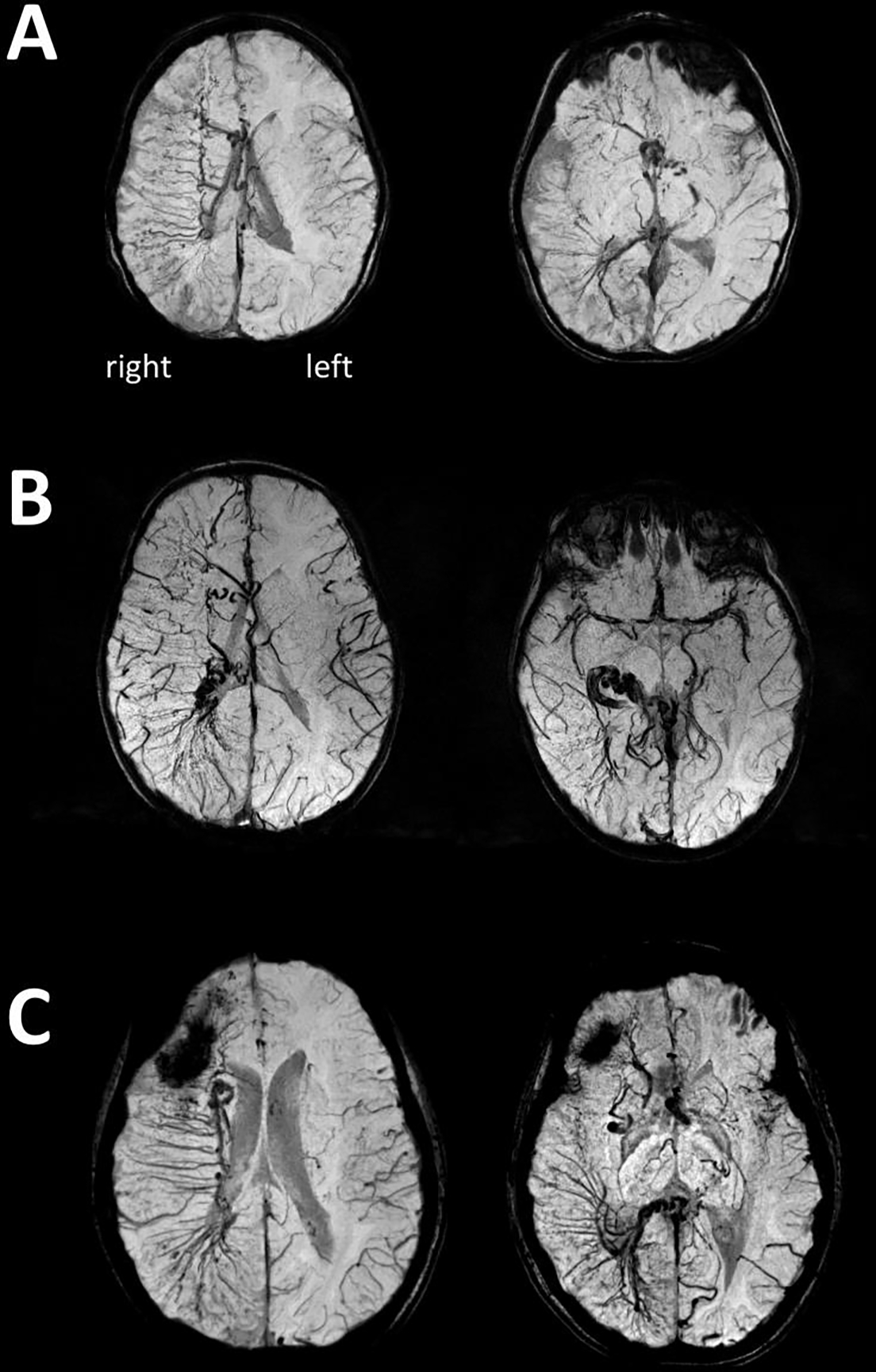
Background: Enlarged deep medullary veins (EDMVs) in patients with Sturge-Weber syndrome (SWS) may provide compensatory venous drainage for brain regions affected by the leptomeningeal venous malformation (LVM). We evaluated the prevalence, extent, hemispheric differences, and clinical correlates of EDMVs in SWS.

Methods: Fifty children (median age: 4.5 years) with unilateral SWS underwent brain magnetic resonance imaging prospectively including susceptibility-weighted imaging (SWI); children aged 2.5 years or older also had a formal neurocognitive evaluation. The extent of EDMVs was assessed on SWI by using an EDMV hemispheric score, which was compared between patients with right and left SWS and correlated with clinical variables.

Results: EDMVs were present in 89% (24 of 27) of right and 78% (18 of 23) of left SWS brains. Extensive EDMVs (score >6) were more frequent in right (33%) than in left SWS (9%; P = 0.046) and commonly occurred in young children with right SWS. Patients with EDMV scores >4 had rare (less than monthly) seizures, whereas 35% (11 of 31) of patients with EDMV scores ≤4 had monthly or more frequent seizures (P = 0.003). In patients with right SWS and at least two LVM-affected lobes, higher EDMV scores were associated with higher intelligence quotient (P < 0.05).

Conclusions: Enlarged deep medullary veins are common in unilateral SWS, but extensive EDMVs appear to develop more commonly and earlier in right hemispheric SWS. Deep venous remodeling may be a compensatory mechanism contributing to better clinical outcomes in some patients with SWS.

Keywords: Cerebral veins; Cognitive functions; Magnetic resonance imaging; Remodeling; Seizures; Sturge-Weber syndrome.


Conflict of interest statement

Conflict of interest: none.

Figures

Figure 1.
Figure 1.
Examples of the extent of enlarged deep medullary veins (EDMVs) in the central (A), frontal (B), parietal (C), and temporal or occipital (D) venous regions. All venous abnormalities are shown in the right hemisphere for an easier comparison, and images from left SWS have been flipped for this purpose. For each image on panels A-C, only one region is framed (red frame in central, orange frame in frontal, yellow frame in parietal regions) even if additional EDMVs are present in other venous regions. On panel D, temporal lobe veins are framed with blue (scores 1–3) and occipital lobe veins with purple (score 1 only).
Figure 2.
Figure 2.
Three examples of extensive right hemispheric enlarged deep veins (EDMVs) as shown by SWI minimum intensity projection images of a: (A) 2.4-year-old child, (B) 4.4-year-old child, and (C) 7.9-year-old child. All three children developed well cognitively and had only rare seizures (frequency score 0 or 1) despite extensive right hemispheric involvement. The first images show frontal, central, and parietal EDMVs, while the second images are from a lower plane showing temporo-occipital and/or inferior frontal EDMVs. Enlarged subependymal veins are also visualized. On panel C, the dark area in the right frontal region corresponded to a calcified region.
Figure 3.
Figure 3.
(A) Age-related increase in enlarged deep medullary vein (EDMV) scores in left hemispheric SWS (Spearman’s rho=0.48, p=0.02). (B) In patients with right SWS, age was not associated with the EDMV scores (rho=0.03, p=0.89); note that 6 patients with right SWS (but none with left SWS) had high EDMV scores (>6) already before 8 years of age.
Figure 4.
Figure 4.
Representative axial T2 (A), FLAIR (B), post-contrast T1 (C), and SWI MIP (D) images of a child with right hemispheric SWS associated with extensive enlarged deep medullary veins (EDMVs). These veins are best visualized by SWI, which also shows the extent of posterior LVM as well as enlarged subependymal veins on the right side. T2 and FLAIR images show atrophy of the affected parietal and frontal lobes and also visualize portions of enlarged subependymal veins and choroid plexus. The post-contrast T1 image demonstrates right parietal leptomeningeal enhancement and also shows some of the enlarged deep medullary veins along with the enlarged choroid plexus in the right hemisphere.
Figure 5.
Figure 5.
Correlations of enlarged deep medullary vein (EDMV) scores with PIQ (A, B) and VIQ (C, D) in patients with at least two LVM-affected lobes. Higher EDMV scores were associated with higher PIQ (rho=0.56, p=0.02) and VIQ (rho=0.52, p=0.03) in right SWS (n=17). The correlations were not significant in left SWS (PIQ: rho=−0.27, p=0.33; VIQ: rho=−0.31, p=0.25; n=15).

References

    1. Ao DH, Zhang DD, Zhai FF, Zhang JT, Han F, Li ML, Ni J, Yao M, Zhang SY, Cui LY, Jin ZY, Zhou LX, Zhu YC. Brain deep medullary veins on 3-T MRI in a population-based cohort. J Cereb Blood Flow Metab. 2021;41:561–568. - PMC - PubMed
    1. Behen ME, Juhász C, Wolfe-Christensen C, Guy W, Halverson S, Rothermel R, Janisse J, Chugani HT. Brain damage and IQ in unilateral Sturge-Weber syndrome: Support for a “fresh start” hypothesis. Epilepsy Behav 2011;22:352–357. - PMC - PubMed
    1. Bentson JR, Wilson GH, Newton TH. Cerebral venous drainage pattern of the Sturge-Weber syndrome. Radiology 1971;101:111–118. - PubMed
    1. Bériault S, Xiao Y, Collins DL, Pike GB. IEEE Trans Med Imaging. Automatic SWI venography segmentation using conditional random fields. 2015;34:2478–2491. - PubMed
    1. Bodensteiner JB, Roach ES. Overview of Sturge-Weber syndrome. In: Bodensteiner JB, Roach ES, eds. Sturge-Weber syndrome. Mt Freedom, NJ: The Sturge-Weber Foundation, 2010:19–32.

Publication types