. 2023 May;50(6):1629-1635.
doi: 10.1007/s00259-022-06081-4. Epub 2022 Dec 16.

Assessment of myocardial fibrosis in patients with systemic sclerosis using [68Ga]Ga-FAPI-04-PET-CT

Affiliations

Assessment of myocardial fibrosis in patients with systemic sclerosis using [68Ga]Ga-FAPI-04-PET-CT

Christoph Treutlein et al. Eur J Nucl Med Mol Imaging. 2023 May.

Abstract

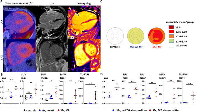
Purpose: Myocardial fibrosis (MF) is a factor of poor prognosis in systemic sclerosis (SSc). Direct in-vivo visualization of fibroblast activation as early readout of MF has not been feasible to date. Here, we characterize 68Gallium-labeled-Fibroblast-Activation-Inhibitor-04 ([68Ga]Ga-FAPI-04)-PET-CT as a diagnostic tool in SSc-related MF.

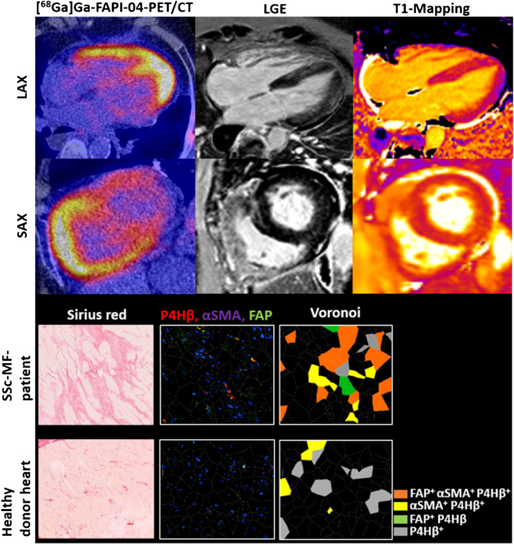
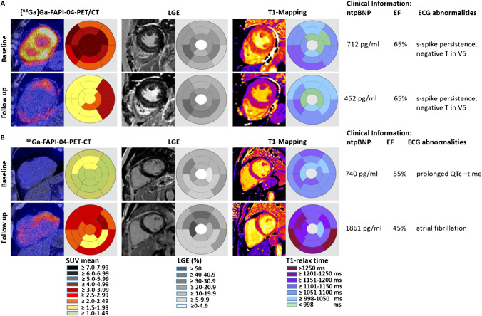
Methods: In this proof-of-concept trial, six SSc patients with and eight without MF of the EUSTAR cohort Erlangen underwent [68Ga]Ga-FAPI-04-PET-CT and cardiac MRI (cMRI) and clinical and serologic investigations just before baseline and during follow-up between January 2020 and December 2020. Myocardial biopsy was performed as clinically indicated.

Results: [68Ga]Ga-FAPI-04 tracer uptake was increased in SSc-related MF with higher uptake in SSc patients with arrhythmias, elevated serum-NT-pro-BNP, and increased late gadolinium enhancement (LGE) in cMRI. Histologically, myocardial biopsies from cMRI- and [68Ga]Ga-FAPI-04-positive regions confirmed the accumulation of FAP+ fibroblasts surrounded by collagen deposits. We observed similar but not equal spatial distributions of [68Ga]Ga-FAPI-04 uptake and quantitative cMRI-based techniques. Using sequential [68Ga]Ga-FAPI-04-PET-CTs, we observed dynamic changes of [68Ga]Ga-FAPI-04 uptake associated with changes in the activity of SSc-related MF, while cMRI parameters remained stable after regression of molecular activity and rather indicated tissue damage.

Conclusions: We present first in-human evidence that [68Ga]Ga-FAPI-04 uptake visualizes fibroblast activation in SSc-related MF and may be a diagnostic option to monitor cardiac fibroblast activity in situ.

Keywords: Cardiac MRI; Myocardial fibrosis; Systemic sclerosis; [68Ga]Ga-FAPI-04-PET-CT.


Conflict of interest statement

JHWD has consultancy relationships and/or has received research funding from Actelion, BMS, Celgene, Bayer Pharma, Boehringer Ingelheim, JB Therapeutics, Sanofi-Aventis, Novartis, UCB, GSK, Array Biopharma, Galapagos, Inventiva, and Active Biotech in the area of potential treatments of SSc and is stock owner of 4D Science. CB has received consultancy fees from Pfizer, Janssen, and Boehringer Ingelheim. KT has received consultancy fees and honoraria from UCB and Gilead. CS has received consultancy fees from ROTOP Pharmaka GmbH and Lilly Pharmaceuticals.

Figures

Fig. 1
Fig. 1
[68Ga]Ga-FAPI-04 uptake is increased in SSc patients with myocardial fibrosis compared to SSc patients without myocardial fibrosis and nondiseased controls. A Representative image of a [68Ga]Ga-FAPI-04-PET/CT scan of a patient with Systemic Sclerosis (SSc)-related myocardial fibrosis (MF) with tracer uptake in fibrotic areas of the myocardium of both ventricles. The corresponding cMRI confirms that [68Ga]Ga-FAPI-04 tracer uptake projects to areas of fibrotic tissue remodeling as visualized by late gadolinium enhancement (LGE) and prolonged T1-relaxation time. B Tissue to background ratio (TBR), maximal and mean standardized uptake values (SUV max and SUV mean (median/IQR)), metabolically active volume (MAV, cm3, (median/IQR)), and total lesion FAPI (TL-FAPI, cm3 (median/IQR)) in patients with SSc-associated MF (n = 6), in SSc patients without MF (n = 8), and in nondiseased controls (n = 5). C Mapping of the SUV mean values to the 17-regions of the AHA-model in patients with SSc-associated MF (n = 6), in SSc patients without MF (n = 8) and in nondiseased controls. Visualization of the mean SUVmean values per group in every segment. The individual distribution of SUVmean values of all SSc patients are shown in supplementary Fig. 2. D TBR, SUV max, SUV mean, MAV (cm3), and TL-FAPI (cm3) of SSc patients with and without ECG-abnormalities (6/8 per group corresponding to the groups with myocardial involvement). Data are presented as the median with an interquartile range. *p < 0.05; **p < 0.01; ***p < 0.001. SAX: short axis view, LAX: long axis view, ECG: electrocardiogram, myocard. dis.: myocardial disease
Fig. 2
Fig. 2
Increased [68Ga]Ga-FAPI-04 uptake corresponds to the accumulation of FAP+-expressing myofibroblasts in myocardial biopsy. Representative image of a [68Ga]Ga-FAPI-04-PET-CT scan and the corresponding image of cardiac MRI with late gadolinium enhancement (LGE) and T1-mapping of a patient with myocardial fibrosis (MF) in the upper part of the image. Myocardial biopsy was performed from a [68Ga]Ga-FAPI-04 positive- and LGE-positive region. Representative pictures of sirius red staining and immunofluorescence staining with the fibroblast marker prolyl-hydroxylase-4β (P4Hβ), the myofibroblast marker α-smooth-muscle actin (αSMA), and fibroblast activation protein (FAP) and Voronoi tessellations are shown in the lower part of the figure. Myocardial tissue sections of two healthy donor hearts of heart-transplanted patients were analyzed as controls. SAX: short axis view, LAX: long axis view
Fig. 3
Fig. 3
Follow-up observations of SSc-MF patients. Representative images of [68Ga]Ga-FAPI-04-PET-CT scan and the corresponding cMRI with late gadolinium enhancement (LGE) and T-mapping at baseline (BL) and follow-up (FU). Clinical findings including serum Nt-pro-BNP levels, ejection fraction (EF, %), and ECG findings are tabulated. A Reduction of [68Ga]Ga-FAPI-tracer uptake upon start of medical therapy in a patient with pronounced tracer uptake at baseline, stable LGE, and minor changes of T1 relaxation times (participant 13). B Progressive [68Ga]Ga-FAPI-04 tracer uptake and clinical deterioration with increased NT-pro-BNP levels and new onset of atrial fibrillation on follow op despite therapy (participant 10). The follow-up investigations of participants 11 and 14 are visualized in supplementary Fig. 6

References

    1. Denton CP, Khanna D. Systemic sclerosis. Lancet. 2017;390(10103):1685–1699. doi: 10.1016/s0140-6736(17)30933-9. - DOI - PubMed
    1. Gabrielli A, Avvedimento EV, Krieg T. Scleroderma. N Engl J Med. 2009;360(19):1989–2003. doi: 10.1056/NEJMra0806188. - DOI - PubMed
    1. Allanore Y, Distler O, Walker UA, Khanna D, Furst DE, Meune C. Points to consider when doing a trial primarily involving the heart. Rheumatology (Oxford). 2017;56(suppl_5):v12-v6. 10.1093/rheumatology/kex198. - PMC - PubMed
    1. Tyndall AJ, Bannert B, Vonk M, Airò P, Cozzi F, Carreira PE, et al. Causes and risk factors for death in systemic sclerosis: a study from the EULAR Scleroderma Trials and Research (EUSTAR) database. Ann Rheum Dis. 2010;69(10):1809–1815. doi: 10.1136/ard.2009.114264. - DOI - PubMed
    1. Meune C, Vignaux O, Kahan A, Allanore Y. Heart involvement in systemic sclerosis: evolving concept and diagnostic methodologies. Arch Cardiovasc Dis. 2010;103(1):46–52. doi: 10.1016/j.acvd.2009.06.009. - DOI - PubMed