. 2022 Dec 15;22(1):607.
doi: 10.1186/s12903-022-02647-8.

Effect of partial restorative treatment on stress distributions in non-carious cervical lesions: a three-dimensional finite element analysis

Affiliations

Effect of partial restorative treatment on stress distributions in non-carious cervical lesions: a three-dimensional finite element analysis

Xin Luo et al. BMC Oral Health. .

Abstract

Background: Partial restoration combined with periodontal root coverage surgery can be applied to the treatment of non-carious cervical lesions (NCCLs) accompanied with gingival recessions in clinical practice. However, the feasibility of NCCL partial restorative treatment from a biomechanical perspective remains unclear. This study aimed to investigate the effect of partial restorations on stress distributions in the NCCLs of mandibular first premolars via three-dimensional finite element analysis.

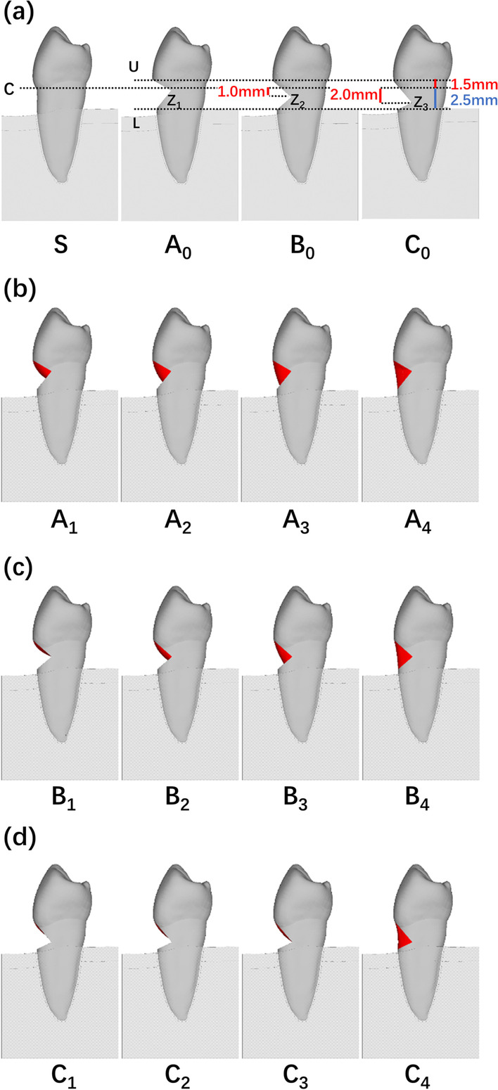
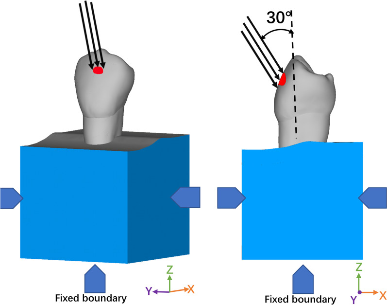
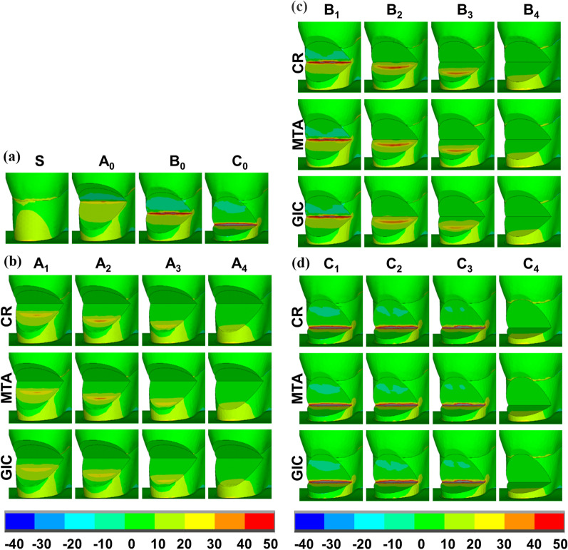
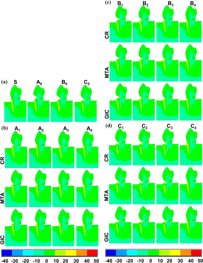
Methods: Three-dimensional finite element models of buccal wedge-shaped NCCLs in various locations of a defected zenith (0 mm, 1 mm, and 2 mm) were constructed and divided into three groups (A, B, and C). Three partially restored NCCL models with different locations of the lower restoration border (1 mm, 1.5 mm, and 2 mm), and one completely restored NCCL model were further constructed for each group. The following restorative materials were used in all restoration models: composite resin (CR), glass-ionomer cement (GIC), and mineral trioxide aggregate (MTA). The first principal stress distributions under buccal oblique loads of 100 N were analyzed. Restoration bond failures were also evaluated based on stress distributions at dentin-restoration interfaces.

Results: When the partial restoration fully covered the defected zenith, the first principal stress around the zenith decreased and the maximum tensile stress was concentrated at the lower restoration border. When the partial restoration did not cover the defected zenith, the first principal stress distribution patterns were similar to those in unrestored models, with the maximum tensile stress remaining concentrated at the zenith. As the elastic modulus of the restorative material was altered, the stress distributions at the interface were not obviously changed. Restoration bond failures were not observed in CR, but occurred in GIC and MTA in most models.

Conclusions: Partial restorations that fully covered defected zeniths improved the stress distributions in NCCLs, while the stress distributions were unchanged or worsened under other circumstances. CR was the optimal material for partial restorations compared to GIC and MTA.

Keywords: Composite resin (CR); Finite element analysis; Glass-ionomer cement (GIC); Mineral trioxide aggregate (MTA); Non-carious cervical lesions (NCCLs); Partial restorations; Restoration bond failures.


Conflict of interest statement

The authors declare that they have no conflict of interest.

Figures

Fig. 1
Fig. 1
Schematic representation of the finite element models. a Detailed parameters of unrestored NCCLs (A0, B0, C0); b partially restored NCCLs (A1–A3) and a completely restored NCCL (A4); c partially restored NCCLs (B1–B3) and a completely restored NCCL (B4); d partially restored NCCLs (C1–C3) and a completely restored NCCL (C4)
Fig. 2
Fig. 2
Schematic representation of the buccal oblique load (the black vector indicates the direction of the applied load. The blue areas indicate the fixed boundary conditions of the model at the lateral and basal surfaces.)
Fig. 3
Fig. 3
Specific regions analyzed for first principal stress (MPa) distributions in models using different restorative materials. a Natural tooth (S) and unrestored NCCLs (A0, B0, C0); b partially restored NCCLs (A1–A3)
Fig. 4
Fig. 4
Close-up view of cervical regions for first principal stress (MPa) distributions in the models where restorations were placed on transparently to enable NCCL wall visualization. a Natural tooth (S) and unrestored NCCLs (A0, B0, C0); b partially restored NCCLs (A1–A3) and completely restored NCCLs (A4); c partially restored NCCLs (B1–B3) and completely restored NCCLs (B4); d partially restored NCCLs (C1–C3) and completely restored NCCLs (C4)
Fig. 5
Fig. 5
Sagittal sections of first principal stress (MPa) distributions in models using different restorative materials. a Natural tooth (S) and unrestored NCCLs (A0, B0, C0); b partially restored NCCLs (A1–A3) and completely restored NCCLs (A4); c partially restored NCCLs (B1–B3) and completely restored NCCLs (B4); d partially restored NCCLs (C1–C3) and completely restored
Fig. 6
Fig. 6
First principal stress and shear stress (MPa) distributions at dentin-restoration interfaces for partial restorations and complete restorations using different restorative materials. a First principal stress distributions in partially restored NCCLs (A1–A3) and completely restored NCCLs (A4); b shear stress distributions in partially restored NCCLs (A1–A3) and completely restored NCCLs (A4); c first principal stress distributions in partially restored NCCLs (B1–B3) and completely restored NCCLs (B4); d shear stress distributions in partially restored NCCLs (B1–B3) and completely restored NCCLs (B4); e first principal stress distributions in partially restored NCCLs C1–C3 and completely restored NCCLs (C4); f shear stress distributions in partially restored NCCLs (C1–C3) and completely restored NCCLs (C4)

References

    1. Teixeira DNR, Thomas RZ, Soares PV, Cune MS, Gresnigt MMM, Slot DE. Prevalence of noncarious cervical lesions among adults: a systematic review. J Dent. 2020;95:103285. doi: 10.1016/j.jdent.2020.103285. - DOI - PubMed
    1. Pini-Prato G, Franceschi D, Cairo F, Nieri M, Rotundo R. Classification of dental surface defects in areas of gingival recession. J Periodontol. 2010;81(6):885–890. doi: 10.1902/jop.2010.090631. - DOI - PubMed
    1. Zucchelli G, Testori T, De Sanctis M. Clinical and anatomical factors limiting treatment outcomes of gingival recession: a new method to predetermine the line of root coverage. J Periodontol. 2006;77(4):714–721. doi: 10.1902/jop.2006.050038. - DOI - PubMed
    1. Zucchelli G, Mounssif I. Periodontal plastic surgery. Periodontol 2000. 2015;68(1):333–368. doi: 10.1111/prd.12059. - DOI - PubMed
    1. Zucchelli G, Gori G, Mele M, Stefanini M, Mazzotti C, Marzadori M, Montebugnoli L, De Sanctis M. Non-carious cervical lesions associated with gingival recessions: a decision-making process. J Periodontol. 2011;82(12):1713–1724. doi: 10.1902/jop.2011.110080. - DOI - PubMed

MeSH terms