. 2022 Dec 15;23(1):349.
doi: 10.1186/s12931-022-02267-4.

Characterization of pulmonary vascular remodeling and MicroRNA-126-targets in COPD-pulmonary hypertension

Affiliations

Characterization of pulmonary vascular remodeling and MicroRNA-126-targets in COPD-pulmonary hypertension

Khushboo Goel et al. Respir Res. .

Abstract

Background: Despite causing increased morbidity and mortality, pulmonary hypertension (PH) in chronic obstructive pulmonary disease (COPD) patients (COPD-PH) lacks treatment, due to incomplete understanding of its pathogenesis. Hypertrophy of pulmonary arterial walls and pruning of the microvasculature with loss of capillary beds are known features of pulmonary vascular remodeling in COPD. The remodeling features of pulmonary medium- and smaller vessels in COPD-PH lungs are less well described and may be linked to maladaptation of endothelial cells to chronic cigarette smoking (CS). MicroRNA-126 (miR126), a master regulator of endothelial cell fate, has divergent functions that are vessel-size specific, supporting the survival of large vessel endothelial cells and inhibiting the proliferation of microvascular endothelial cells. Since CS decreases miR126 in microvascular lung endothelial cells, we set out to characterize the remodeling by pulmonary vascular size in COPD-PH and its relationship with miR126 in COPD and COPD-PH lungs.

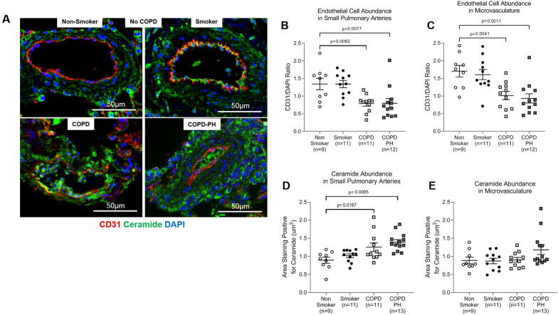
Methods: Deidentified lung tissue was obtained from individuals with COPD with and without PH and from non-diseased non-smokers and smokers. Pulmonary artery remodeling was assessed by ⍺-smooth muscle actin (SMA) abundance via immunohistochemistry and analyzed by pulmonary artery size. miR126 and miR126-target abundance were quantified by qPCR. The expression levels of ceramide, ADAM9, and endothelial cell marker CD31 were assessed by immunofluorescence.

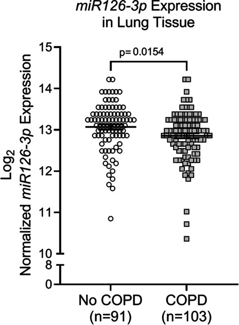
Results: Pulmonary arteries from COPD and COPD-PH lungs had significantly increased SMA abundance compared to non-COPD lungs, especially in small pulmonary arteries and the lung microvasculature. This was accompanied by significantly fewer endothelial cell markers and increased pro-apoptotic ceramide abundance. miR126 expression was significantly decreased in lungs of COPD individuals. Of the targets tested (SPRED1, VEGF, LAT1, ADAM9), lung miR126 most significantly inversely correlated with ADAM9 expression. Compared to controls, ADAM9 was significantly increased in COPD and COPD-PH lungs, predominantly in small pulmonary arteries and lung microvasculature.

Conclusion: Both COPD and COPD-PH lungs exhibited significant remodeling of the pulmonary vascular bed of small and microvascular size, suggesting these changes may occur before or independent of the clinical development of PH. Decreased miR126 expression with reciprocal increase in ADAM9 may regulate endothelial cell survival and vascular remodeling in small pulmonary arteries and lung microvasculature in COPD and COPD-PH.

Keywords: ADAM9; Chronic obstructive pulmonary disease; Emphysema; Endothelial cells; Group 3 pulmonary hypertension; Pulmonary vascular remodeling; Smoking; microRNA-126.


Conflict of interest statement

The authors declare that they have no competing interests.

Figures

Fig. 1
Fig. 1
Pulmonary Arterial Remodeling in COPD and COPD-PH. A Representative images of smooth muscle actin (SMA) (brown) in human lung tissue from non-smokers and smokers without COPD as well as COPD and COPD-PH individuals, determined by immunohistochemistry (IHC). The slides were counterstained with hematoxylin (blue). Black arrows point to pulmonary arteries. Images were captured at 10X; scale bar is 100 µm. B–E Quantification of pulmonary arterial remodeling was performed by calculating the proportion of the artery which stained positive for SMA. B Quantification of SMA abundance in pulmonary arteries, regardless of size. Each data point represents an individual; horizontal lines are mean ± SEM. SMA abundance analyzed by size of: (C) medium pulmonary arteries, (D) small pulmonary arteries, and (E) microvasculature, with each data point representing a single pulmonary vessel and horizontal lines representing mean ± SD. For all graphs, n = number of individuals in each group; 1-way ANOVA with Dunnett’s multiple comparisons test was used for statistical analysis
Fig. 2
Fig. 2
Endothelial cell and ceramide abundance in pulmonary arteries. A Representative images of immunofluorescence performed on human lung tissue from non-smokers and smokers without COPD as well as COPD and COPD-PH individuals, staining for the endothelial cell marker CD31 (red), ceramide (green), and DAPI (blue). Each image shows a pulmonary artery. Images were captured at 20×; scale bar is 50 µm. B, C Quantification of endothelial cell abundance in (B) small pulmonary arteries and (C) the microvasculature. D, E Quantification of ceramide abundance in (D) small pulmonary arteries and (E) the microvasculature. For all graphs, each data point represents an individual; n = number of individuals in each group; horizontal lines are mean ± SEM; 1-way ANOVA with Dunnett’s multiple comparisons test was used for statistical analysis
Fig. 3
Fig. 3
miR126 Expression in COPD. MiR126-3p expression from reanalysis of microarray profiles of human lung tissue from individuals with or without COPD performed by the Lung Genomics Research Consortium (LGRC). The samples were obtained from the NHLBI-sponsored Lung Tissue Research Consortium (LTRC) (GSE72967, GSE47460). Each data point represents an individual; horizontal lines are mean ± SEM; unpaired t-test was used for statistical analysis
Fig. 4
Fig. 4
Correlation between miR126 and ADAM9. A Linear regression of association of lung miR126 and ADAM9 mRNA expression levels measured by RTqPCR in non-smokers (n = 4), smokers without COPD (n = 3), and COPD-PH (n = 1) individuals. Each data point represents an individual; Pearson’s correlation analysis was used for statistical analysis. B ADAM9 long (L) and short (S) isoforms abundance in western blots of human lung microvascular endothelial cells from non-smokers transfected with miR126-3p-mimic (126-OE) or antisense (126-KD), using vinculin as loading control. Below, normalized levels are quantified by densitometry
Fig. 5
Fig. 5
ADAM9 abundance in pulmonary arteries. A Representative images of ADAM9 (Red) endothelial cell marker CD31 (green), and DAPI (blue) abundance detected by immunofluorescence performed on human lung tissue from non-smokers and smokers without COPD, GOLD stage 4 COPD, and COPD-PH individuals. White arrows point to pulmonary arteries. Images were captured at 20X; scale bar is 50 µm. B, C Quantification of ADAM9 abundance in (B) small pulmonary arteries and (C) the microvasculature. For all graphs, each data point represents an individual; n = number of individuals in each group; horizontal lines are mean ± SEM; 1-way ANOVA with Dunnett’s multiple comparisons test was used for statistical analysis

References

    1. Tuder RM, Petrache I. Pathogenesis of chronic obstructive pulmonary disease. J Clin Invest. 2012;122(8):2749–2755. doi: 10.1172/JCI60324. - DOI - PMC - PubMed
    1. Cornelius ME, Wang TW, Jamal A, Loretan CG, Neff LJ. Tobacco product use among adults—United States, 2019. Morb Mortal Wkly Rep. 2020;69(46):1736–1742. doi: 10.15585/mmwr.mm6946a4. - DOI - PMC - PubMed
    1. Petrache INV, Zhen L, Medler TR, Richter A, Cho C, Hubbard WC, Berdyshev EV, Tuder RM. Ceramide upregulation causes pulmonary cell apoptosis and emphysema. Nat Med. 2005;11(5):491–498. doi: 10.1038/nm1238. - DOI - PMC - PubMed
    1. Minai OA, Chaouat A, Adnot S. Pulmonary hypertension in COPD: epidemiology, significance, and management: pulmonary vascular disease: the global perspective. Chest. 2010;137(6 Suppl):39S–51S. doi: 10.1378/chest.10-0087. - DOI - PubMed
    1. Oswald-Mammosser M, Weitzenblum E, Quoix E, Moser G, Chaouat A, Charpentier C, et al. Prognostic factors in COPD patients receiving long-term oxygen therapy. Importance of pulmonary artery pressure. Chest. 1995;107(5):1193–1198. doi: 10.1378/chest.107.5.1193. - DOI - PubMed

MeSH terms