

Hemoperitoneum, Hepatic Laceration, and Hepatic Artery Pseudoaneurysm as a Complication of Emergent Pericardiocentesis
- PMID: 36523950
- PMCID: PMC9745653
- DOI: 10.1016/j.jaccas.2022.101686
Hemoperitoneum, Hepatic Laceration, and Hepatic Artery Pseudoaneurysm as a Complication of Emergent Pericardiocentesis
Abstract
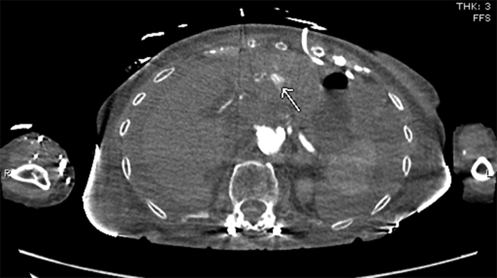
Emergent pericardiocentesis is a potentially life-saving therapeutic procedure. We report a case of hemoperitoneum, a rare but known complication of pericardiocentesis; due to hepatic artery laceration and hepatic artery pseudoaneurysm formation resulting in delayed hemorrhagic shock as a complication of emergent pericardiocentesis. (Level of Difficulty: Intermediate.).
Keywords: CTA, computed tomography angiogram; LA, left atrium; MR, mitral regurgitation; TEE, transesophageal echocardiogram; TEER, transcatheter mitral valve edge-to-edge repair; TTE, transthoracic echocardiogram; hemoperitoneum; hepatic artery laceration; pericardial tamponade; pericardiocentesis.
© 2022 The Authors.
Conflict of interest statement
The authors have reported that they have no relationships relevant to the contents of this paper to disclose.
Figures


Comment in
-
Removing the Blindfold: Echo-Guidance for Pericardiocentesis.JACC Case Rep. 2022 Dec 12;5:101699. doi: 10.1016/j.jaccas.2022.101699. eCollection 2023 Jan 4. JACC Case Rep. 2022. PMID: 36636510 Free PMC article.
References
-
- Adler Y., Charron P., Imazio M., et al. 2015 ESC guidelines for the diagnosis and management of pericardial diseases: The task force for the diagnosis and management of pericardial diseases of the European Society of Cardiology (ESC) endorsed by the European Association for Cardio-Thoracic Surgery (EACTS) Eur Heart J. 2015;36 - PubMed
-
- Jareño Martínez S., Bruna Esteban M., Núñez Ronda R., Grifo Albalat I., Fabregat-Andrés Ó Hemoperitoneum due to left inferior phrenic artery injury during pericardiocentesis. Rev Esp Cardiol (Engl Ed) 2015;68:1031–1032. - PubMed
Publication types
LinkOut - more resources
Full Text Sources
