. 2022 Dec 16;8(50):eabn6025.
doi: 10.1126/sciadv.abn6025. Epub 2022 Dec 16.

β-Endorphin mediates radiation therapy fatigue

Affiliations

β-Endorphin mediates radiation therapy fatigue

Andrea L Hermann et al. Sci Adv. .

Abstract

Fatigue is a common adverse effect of external beam radiation therapy in cancer patients. Mechanisms causing radiation fatigue remain unclear, although linkage to skin irradiation has been suggested. β-Endorphin, an endogenous opioid, is synthesized in skin following genotoxic ultraviolet irradiation and acts systemically, producing addiction. Exogenous opiates with the same receptor activity as β-endorphin can cause fatigue. Using rodent models of radiation therapy, exposing tails and sparing vital organs, we tested whether skin-derived β-endorphin contributes to radiation-induced fatigue. Over a 6-week radiation regimen, plasma β-endorphin increased in rats, paralleled by opiate phenotypes (elevated pain thresholds, Straub tail) and fatigue-like behavior, which was reversed in animals treated by the opiate antagonist naloxone. Mechanistically, all these phenotypes were blocked by opiate antagonist treatment and were undetected in either β-endorphin knockout mice or mice lacking keratinocyte p53 expression. These findings implicate skin-derived β-endorphin in systemic effects of radiation therapy. Opioid antagonism may warrant testing in humans as treatment or prevention of radiation-induced fatigue.


Figures

Fig. 1.
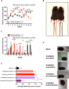
Fig. 1.. Tail radiation increases β-endorphin levels, depresses motor activity, and induces other opiate phenotypes in rats.
Sprague-Dawley rats underwent tail radiation or mock treatment (2 Gy/day) 5 days/week for 6 weeks. Error bars in the figure indicate ±SEM. Numbers in brackets indicate numbers of animals per group. Arrows in (A) and (B) indicate the last day of radiation exposure or mock treatment (week 6, day 42). In (A) and (B), 6 weeks of tail radiation (5 days/week) or mock exposure were done on all groups followed by two additional weeks after tail treatment. (A) Plasma β-endorphin levels for each of the groups. Two-way analysis of variance (ANOVA) with the Holm-Šídák multiple comparisons test revealed significant differences between groups that are indicated above the graphs. Black numbers indicate differences between Mock-Saline and Irradiated-Saline; blue numbers indicate differences between Mock-Naloxone and Irradiated-Naloxone groups. (B) Distance traveled in 30 min by open-field actimetry was measured once weekly as described in (A). Rats were administered intraperitoneal saline injection or naloxone injection (10 mg/kg) before actimetry testing. Two-way ANOVA with the Holm-Šídák multiple comparisons test revealed significant differences between groups that are indicated above the graphs. Red numbers indicate difference between Irradiated-Naloxone and Irradiated-Saline. Black number indicates difference between Irradiated-Saline and Mock-Saline. All other groups showed no significant change from baseline. (C) Straub tail reaction is observed after 5 weeks of tail radiation (bottom), but not in mock-irradiated animals (top). Value of significance was determined by two-tailed unpaired t test. (D) Thermal nociceptive threshold increased in irradiated animals. Two-way ANOVA with the Holm-Šídák multiple comparisons test revealed significant differences between the groups that are indicated above the graphs. Black numbers indicate differences between Mock and Irradiated rats.
Fig. 2.
Fig. 2.. Tail ionizing radiation exposure in mice increases plasma β-endorphin levels and induces opioid-dependent Straub tail.
(A) Weekly plasma β-endorphin levels in wild-type mice in a crossover experiment in which, initially, one group of mice underwent mock irradiation, while a second group of mice underwent tail radiation (5 Gy/day) 5 days/week for 6 weeks. After week 6 (day 42), the groups switched treatment regimens, and the treatments continued for six more weeks. Error bars indicate ±SEM. Two-way ANOVA with the Holm-Šídák multiple comparisons test revealed significant differences between the groups that are indicated above the graph. Black numbers indicate differences between Radiation then Mock and Mock then Radiation. (B) Increased local pigmentation of the tail (radiation-exposed area) in a tail-irradiated mouse after 6 weeks of ionizing radiation (left), while no pigmentation is observed on the tail of a mock-treated mouse (right). (C) Straub tail scores in mock-irradiated mice and mice administered tail radiation (5 Gy/day) 5 days/week for 6 weeks. After 6 weeks (day 42), the groups switched regimens [the same regimens as in (A) but with separate groups of mice]. Two-way ANOVA with the Holm-Šídák multiple comparisons test revealed significant differences between the groups that are indicated above the graph. (D) Naloxone reverses Straub tail induced by tail irradiation. Representative photos of mice after tail irradiation or mock treatment and 20 min after saline or naloxone injection described in (E). (E) On week 4 of tail irradiation, mice were administered intraperitoneal saline or naloxone (10 mg/kg), and Straub tail scores were measured 20 min after administration. One-way ANOVA with the Holm-Šídák multiple comparisons test revealed significant differences between groups that are indicated above the graph. Error bars in this figure indicate ±SEM. ns, not significant.
Fig. 3.
Fig. 3.. Increased analgesic thresholds induced by tail irradiation are reversed by opioid antagonists.
After week 6 of tail irradiation or mock exposure, mice were assayed for two additional weeks after tail treatment. Arrows indicate the final day of radiation or mock exposure for each group (Mock or Radiation Stop). Two-way ANOVA with the Holm-Šídák multiple comparisons test revealed significant differences between groups that are indicated above the graphs. Black numbers indicate difference between Irradiated-Saline and Mock-Saline groups. Purple numbers indicate differences between Irradiated-Saline and Irradiated-Naloxone groups. Error bars in the figure indicate ±SEM. (A) Mechanical (von Frey assay) (top) and (B) thermal (hot plate assay) (middle) analgesic thresholds in mice that were tail-irradiated (5 Gy/day ionizing radiation) or mock-treated 5 days/week for 6 weeks and administered intraperitoneal injection of either saline or naloxone (10 mg/kg) before analgesic testing. (C) Plasma β-endorphin levels of mice described in (A) and (B).
Fig. 4.
Fig. 4.. Radiation-induced Straub tail and elevated nociceptive thresholds depend on β-endorphin and on keratinocyte-specific p53 expression.
(A) Mechanical (von Frey assay) and (B) thermal (hot plate assay) analgesic thresholds in β-endorphin wild-type and β-endorphin–null mice over 5 weeks of tail radiation (5 Gy/day) 5 days/week. (C) Straub tail was observed starting 3 weeks after irradiation in β-endorphin wild-type mice, but not in mock-treated or β-endorphin–null mice. (D) β-Endorphin and (E) thermal analgesic threshold (hot plate assay) elevations are absent in p53fl/fl mice expressing Cre under the keratinocyte-specific promoter K14 but are present in p53fl/fl mice with no Cre. Mice were treated weekly with tail radiation (5 Gy/day) 5 days/week. (F) Straub tail was absent in K14Cre;p53fl/fl mice, but was present in p53fl/fl mice after 3 and 4 weeks of the tail radiation regimen described in (D) and (E). Error bars in the figure indicate ±SEM. Two-way ANOVA with the Holm-Šídák multiple comparisons tests revealed significant differences between the groups that are indicated above the graphs.

References

    1. Stasi R., Abriani L., Beccaglia P., Terzoli E., Amadori S.,Cancer-related fatigue. Cancer 98,1786–1801 (2003). - PubMed
    1. Greenberg D. B., Sawicka J., Eisenthal S., Ross D.,Fatigue syndrome due to localized radiation. J. Pain Symptom Manage. 7,38–45 (1992). - PubMed
    1. Jacobsen P. B., Thors C. L.,Fatigue in the radiation therapy patient: Current management and investigations. Semin. Radiat. Oncol. 13,372–380 (2003). - PubMed
    1. Hickok J. T., Morrow G. R., Roscoe J. A., Mustian K., Okunieff P.,Occurrence, severity, and longitudinal course of twelve common symptoms in 1129 consecutive patients during radiotherapy for cancer. J. Pain Symptom Manage. 30,433–442 (2005). - PubMed
    1. Back M., Ahern V., Delaney G., Graham P., Steigler A., Wratten C.; New South Wales Breast Radiation Oncology Group ,Absence of adverse early quality of life outcomes of radiation therapy in breast conservation therapy for early breast cancer. Australas. Radiol. 49,39–43 (2005). - PubMed