. 2022 Dec 16;8(50):eabn7983.
doi: 10.1126/sciadv.abn7983. Epub 2022 Dec 16.

EGFR is a master switch between immunosuppressive and immunoactive tumor microenvironment in inflammatory breast cancer

Affiliations

EGFR is a master switch between immunosuppressive and immunoactive tumor microenvironment in inflammatory breast cancer

Xiaoping Wang et al. Sci Adv. .

Abstract

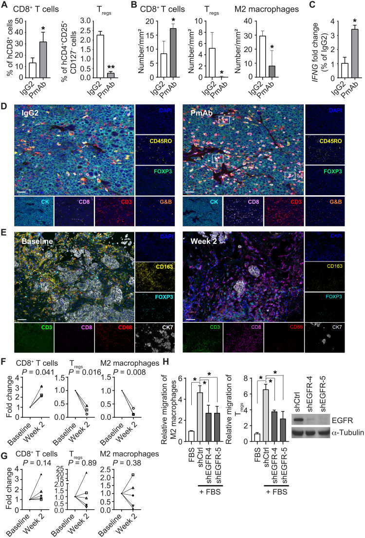
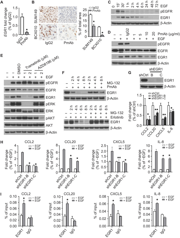
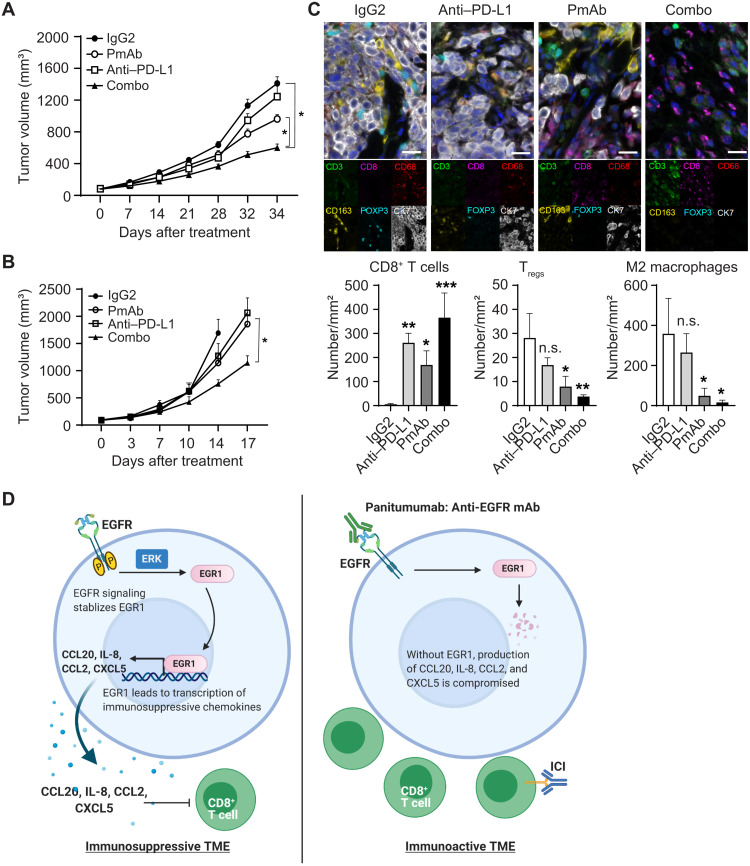
Inflammatory breast cancer (IBC), the most aggressive breast cancer subtype, is driven by an immunosuppressive tumor microenvironment (TME). Current treatments for IBC have limited efficacy. In a clinical trial (NCT01036087), an anti-EGFR antibody combined with neoadjuvant chemotherapy produced the highest pathological complete response rate ever reported in patients with IBC having triple-negative receptor status. We determined the molecular and immunological mechanisms behind this superior clinical outcome. Using novel humanized IBC mouse models, we discovered that EGFR-targeted therapy remodels the IBC TME by increasing cytotoxic T cells and reducing immunosuppressive regulatory T cells and M2 macrophages. These changes were due to diminishing immunosuppressive chemokine expression regulated by transcription factor EGR1. We also showed that induction of an immunoactive IBC TME by an anti-EGFR antibody improved the antitumor efficacy of an anti-PD-L1 antibody. Our findings lay the foundation for clinical trials evaluating EGFR-targeted therapy combined with immune checkpoint inhibitors in patients with cancer.


Figures

Fig. 1.
Fig. 1.. Panitumumab treatment remodels the TME in IBC humanized mouse models.
(A) Tumor growth curve of SUM149 xenograft in humanized mice (SUM149-hu-NSG-SGM3) treated with control IgG2 and panitumumab (five mice per group). Data are summarized as means ± SEM. *P < 0.01. (B) Immunohistochemistry (IHC) staining (left) of phosphorylated EGFR (pEGFR) and Ki67 and quantification of staining intensity (right) in tumor tissues from IgG2- and panitumumab-treated SUM149-hu-NSG-SGM3 mice. *P < 0.01. (C) Tumor growth curve of BCX010 xenograft in humanized mice (BCX010-hu-NSG-SGM3) treated with control IgG2 and panitumumab (six mice per group). Data are summarized as means ± SEM. *P < 0.05. (D) IHC staining (left) of pEGFR and Ki67 and quantification of staining intensity (right) in tumor tissues from IgG2- and panitumumab-treated BCX010-hu-NSG-SGM3 mice. *P < 0.01. (E) Uniform manifold approximation and projection (UMAP) plot of all tumor-resident cells from SUM149-hu-NSG-SGM3 tumors treated with IgG2 and panitumumab (two tumor samples per group). Only cells containing at least 100 gene features and mitochondrial gene counts of less than 20% were used (13,931 cells in IgG2-treated tumor samples and 11,593 cells in panitumumab-treated tumor samples). Clusters denoted by color are labeled with the inferred cell types. (F) Heatmap of pooled gene expression within the epithelial cells among samples treated with IgG2 and panitumumab. These genes are involved in the immune response. (G) Enrichment of pathways associated with epithelial-mesenchymal transition, TNF-α signaling via NF-κB, inflammatory response, IL-6/JAK/STAT3 signaling, and IL-2/STAT5 signaling in IgG2-treated compared to panitumumab-treated SUM149 epithelial tumor cells in humanized mice. (H) Volcano plot of significant differentially expressed genes in CD8+ T cells after panitumumab treatment in SUM149-hu-NSG-SGM3 tumors. PmAb, panitumumab; RBC, red blood cells; FC, fold change; n.s., not significant; FDR, false discover rate.
Fig. 2.
Fig. 2.. Panitumumab treatment affects the TME in IBC tumors.
(A and B) Changes in CD8+ T cells, Tregs, and M2 macrophages in tissues of IgG2- and panitumumab-treated SUM149-hu-NSG-SGM3 mice analyzed by flow cytometry (A) and multiplexed immunofluorescence staining (B). (A) *P = 0.05 and **P < 0.001. (B) *P < 0.05. (C) Panitumumab-treated tissues have increased IFNG gene expression compared to IgG2-treated tissues in SUM149-hu-NSG-SGM3 mice. *P < 0.05. (D) Panitumumab-treated tissues have more CD3+CD8+G&B+ cells than IgG2-treated tissues in SUM149-hu-NSG-SGM3 mice as analyzed by multiplexed immunofluorescence staining. Scale bars, 50 μm. (E to G) Changes in CD8+ T cells, M2 macrophages, and Tregs after panitumumab treatment in matched tissues from three patients with IBC having a pCR (F) or five patients with IBC without a pCR (G) to panitumumab/NAC in primary HER2-negative IBC (NCT01036087). (E) Representative images of multiplexed immunofluorescence staining of CD3, CD8, CD68, CD163, FOXP3, and CK7 in IBC patient tissues at baseline (before panitumumab treatment, left) and week 2 (after panitumumab treatment, right). The numbers of CD8+ T cells (CD3+CD8+), M2 macrophages (CD68+CD163+), and Tregs (CD3+FOXP3+) in five randomly selected areas of each slide were calculated. Each symbol represents the same patient. Scale bars, 50 μm. (H) EGFR knockdown SUM149 clones shEGFR-4 and shEGFR-5 induce less migration of M2 macrophages and Tregs than control knockdown clone shCtrl in in vitro coculture transwell assays. *P < 0.01. G&B, granzyme B; DAPI, 4′,6-diamidino-2-phenylindole; FBS, fetal bovine serum. Experiments in (C) and (H) were independently repeated with three replicates each time.
Fig. 3.
Fig. 3.. Panitumumab treatment affects the TME by regulating chemokine expression in IBC cells.
(A) Expression of chemokines in SUM149-hu-NSG-SGM3 tumors treated with IgG2 and panitumumab by quantitative RT-PCR (qRT-PCR). Three tumor samples per group. *P < 0.01. (B and C) EGFR knockdown (B) or panitumumab treatment (C) affects chemokine expression in SUM149 cells by qRT-PCR. *P < 0.005 and **P < 0.05. (D) Recombinant proteins CCL4, CCL5, CXCL9, and CXCL10 individually or in combination increase the migration of CD8+ T cells. *P < 0.01 and **P < 0.05. (E) Kaplan-Meier metastasis-free survival curves in patients with IBC according to high (N = 70) and low (N = 51) metagene scores of CCL4, CCL5, CXCL9, and CXCL10 mRNA expression. P = 2.62 × 10⁻². (F) Box plot of metagene score of CCL2/CCL20/CXCL5/IL-8 mRNA expression according to 252 non-IBC patients and 137 patients with IBC. P = 5.51 × 10⁻³. (G) Kaplan-Meier overall survival curves in patients with IBC according to high and low CCL20 mRNA expression. P = 2.36 × 10⁻². (H) Recombinant protein CCL20 reduces the migration of CD8+ T cells. *P < 0.05. (I) Recombinant proteins CCL2, CCL20, CXCL5, and IL-8 individually or in combination induce the migration of M2 macrophages. *P < 0.001 and **P < 0.01. (J) Recombinant proteins CCL2 and CCL20 induce the migration of Tregs. *P < 0.01. (D) and (H) to (J) were transwell migration assays. Data are summarized as means ± SD in (A) to (D) and (H) to (J). Experiments in (A) to (D) and (H) to (J) were independently repeated three times with three replicates each time.
Fig. 4.
Fig. 4.. The EGFR pathway regulates the expression of CCL2, CCL20, CXCL5, and IL-8 through EGR1 in IBC.
(A) Panitumumab treatment reduces the gene expression of EGR1 in SUM149 cells. *P < 0.001. (B) Panitumumab treatment reduces the expression of EGR1 protein in SUM149-hu-NSG-SGM3 and BCX010-hu-NSG-SGM3 mice. Left: Representative images. Right: Quantitative data. *P < 0.001. (C) EGF (20 ng/ml) stimulates the expression of EGR1 in SUM149 cells. (D) Pretreatment with panitumumab mitigates the up-regulation of EGR1 by EGF stimulation in SUM149 cells. (E) Pretreatment with MEK inhibitor trametinib, but not PI3K inhibitor AZD8186, inhibits the up-regulation of EGR1 by EGF stimulation in SUM149 cells. (F) MG-132 (5 μM) treatment mitigates the decrease of EGR1 expression induced by panitumumab (20 μg/ml; top) and erlotinib (1 μM; bottom) treatments in SUM149 cells. (G) EGR1 knockdown reduces the expression of CCL2, CCL20, CXCL5, and IL-8 genes in SUM149 cells. *P < 0.05 and **P < 0.01. (H) Effect of EGF stimulation on the expression of CCL2, CCL20, CXCL5, and IL-8 genes in shCtrl and shEGR1-B and shEGR1-C clones of SUM149 cells. P value of shEGR1 clones compared to shCtrl upon EGF stimulation: *P < 0.001 and ** P = 0.01. (I) EGR1 binds to the promoter region of CCL2, CCL20, CXCL5, and IL-8 genes in SUM149 cells upon EGF stimulation. * P < 0.05. Data are summarized as means ± SD in (A) and (G) to (I). DMSO, dimethyl sulfoxide. Experiments were independently repeated three times with three replicates each time.
Fig. 5.
Fig. 5.. Synergistic antitumor effect of panitumumab combined with anti–PD-L1 antibody in humanized mouse models.
(A) Tumor growth curves of SUM149 xenograft in humanized mice (SUM149-hu-NSG-SGM3) treated with control IgG2, panitumumab, anti–PD-L1 antibody, and combination. Eight mice per group. *P < 0.05. (B) Tumor growth curves of BCX010 xenograft in humanized mice (BCX010-hu-NSG-SGM3) treated with control IgG2, panitumumab, anti–PD-L1 antibody, and combination. Eight mice per group. *P < 0.001. (C) Changes in CD8+ T cells, Tregs, and M2 macrophages in tissues of each group in SUM149-hu-NSG-SGM3 mice were analyzed by multiplexed immunofluorescence staining. Scale bars, 50 μm. *P < 0.05, **P < 0.01, and ***P < 0.001. (D) Proposed mechanism by which panitumumab treatment remodels the IBC TME. Left: The activation of EGFR signaling stabilizes EGR1, which regulates the expression of chemokines CCL2, CCL20, CXCL5, and IL-8. The secretion of these chemokines creates an immunosuppressive TME, which attracts Tregs and M2 macrophages but inhibits the recruitment of T cells. Right: Panitumumab inhibits EGFR signaling and induces EGR1 degradation, which reduces the secretion of CCL2, CCL20, CXCL5, and IL-8 from IBC cells, changing the TME from immunosuppressive to immunoactive. Data are summarized as means ± SEM in (A) and (B) and means ± SD in (C).

References

    1. P. Schmid, J. Cortes, L. Pusztai, H. McArthur, S. Kummel, J. Bergh, C. Denkert, Y. H. Park, R. Hui, N. Harbeck, M. Takahashi, T. Foukakis, P. A. Fasching, F. Cardoso, M. Untch, L. Jia, V. Karantza, J. Zhao, G. Aktan, R. Dent, J. O’Shaughnessy; KEYNOTE-522 Investigators ,Pembrolizumab for early triple-negative breast cancer. N. Engl. J. Med. 382, 810–821 (2020). - PubMed
    1. E. A. Mittendorf, H. Zhang, C. H. Barrios, S. Saji, K. H. Jung, R. Hegg, A. Koehler, J. Sohn, H. Iwata, M. L. Telli, C. Ferrario, K. Punie, F. Penault-Llorca, S. Patel, A. N. Duc, M. Liste-Hermoso, V. Maiya, L. Molinero, S. Y. Chui, N. Harbeck,Neoadjuvant atezolizumab in combination with sequential nab-paclitaxel and anthracycline-based chemotherapy versus placebo and chemotherapy in patients with early-stage triple-negative breast cancer (IMpassion031): A randomised, double-blind, phase 3 trial. Lancet 396, 1090–1100 (2020). - PubMed
    1. L. Gandhi, D. Rodriguez-Abreu, S. Gadgeel, E. Esteban, E. Felip, F. De Angelis, M. Domine, P. Clingan, M. J. Hochmair, S. F. Powell, S. Y. Cheng, H. G. Bischoff, N. Peled, F. Grossi, R. R. Jennens, M. Reck, R. Hui, E. B. Garon, M. Boyer, B. Rubio-Viqueira, S. Novello, T. Kurata, J. E. Gray, J. Vida, Z. Wei, J. Yang, H. Raftopoulos, M. C. Pietanza, M. C. Garassino; KEYNOTE-189 Investigators ,Pembrolizumab plus chemotherapy in metastatic non-small-cell lung cancer. N. Engl. J. Med. 378, 2078–2092 (2018). - PubMed
    1. L. Paz-Ares, A. Luft, D. Vicente, A. Tafreshi, M. Gumus, J. Mazieres, B. Hermes, F. Cay Senler, T. Csoszi, A. Fulop, J. Rodriguez-Cid, J. Wilson, S. Sugawara, T. Kato, K. H. Lee, Y. Cheng, S. Novello, B. Halmos, X. Li, G. M. Lubiniecki, B. Piperdi, D. M. Kowalski; KEYNOTE-407 Investigators ,Pembrolizumab plus chemotherapy for squamous non-small-cell lung cancer. N. Engl. J. Med. 379, 2040–2051 (2018). - PubMed
    1. C. Robert, G. V. Long, B. Brady, C. Dutriaux, M. Maio, L. Mortier, J. C. Hassel, P. Rutkowski, C. McNeil, E. Kalinka-Warzocha, K. J. Savage, M. M. Hernberg, C. Lebbe, J. Charles, C. Mihalcioiu, V. Chiarion-Sileni, C. Mauch, F. Cognetti, A. Arance, H. Schmidt, D. Schadendorf, H. Gogas, L. Lundgren-Eriksson, C. Horak, B. Sharkey, I. M. Waxman, V. Atkinson, P. A. Ascierto,Nivolumab in previously untreated melanoma without BRAF mutation. N. Engl. J. Med. 372, 320–330 (2015). - PubMed