

State-of-the-art imaging of neuromodulatory subcortical systems in aging and Alzheimer's disease: Challenges and opportunities
- PMID: 36526031
- PMCID: PMC9805533
- DOI: 10.1016/j.neubiorev.2022.104998
State-of-the-art imaging of neuromodulatory subcortical systems in aging and Alzheimer's disease: Challenges and opportunities
Abstract
Primary prevention trials have shifted their focus to the earliest stages of Alzheimer's disease (AD). Autopsy data indicates that the neuromodulatory subcortical systems' (NSS) nuclei are specifically vulnerable to initial tau pathology, indicating that these nuclei hold great promise for early detection of AD in the context of the aging brain. The increasing availability of new imaging methods, ultra-high field scanners, new radioligands, and routine deep brain stimulation implants has led to a growing number of NSS neuroimaging studies on aging and neurodegeneration. Here, we review findings of current state-of-the-art imaging studies assessing the structure, function, and molecular changes of these nuclei during aging and AD. Furthermore, we identify the challenges associated with these imaging methods, important pathophysiologic gaps to fill for the AD NSS neuroimaging field, and provide future directions to improve our assessment, understanding, and clinical use of in vivo imaging of the NSS.
Keywords: (Functional) magnetic resonance imaging; Alzheimer’s Disease; Brain aging; Diffusion-weighted imaging; Electrophysiology; Neuroimaging; Neuromodulators; Positron emission tomography.
Copyright © 2022 Elsevier Ltd. All rights reserved.
Figures
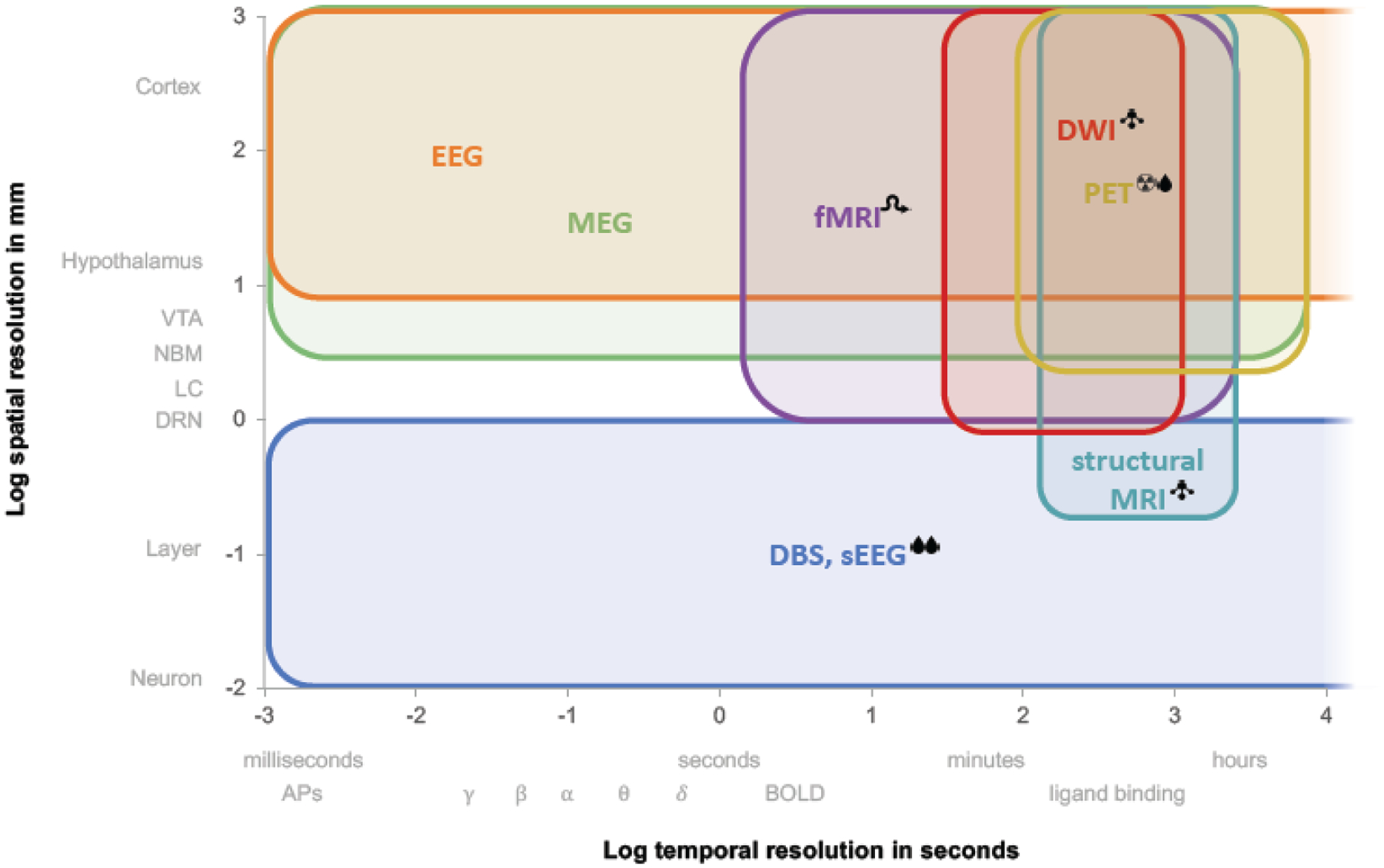
 , invasive;
  , radioactive;
  , structural only;
  , indirect measure of neuronal activity; δ θ α β γ, EEG bands. Abbreviations: APs, action potentials; BOLD, blood oxygen dependent level; DBS, deep brain stimulation; DWI, diffusion-weighted imaging; EEG, electroencephalogram; fMRI, functional magnetic resonance imaging; MEG, magnetoencephalogram; MRI, magnetic resonance imaging; PET, positron emission tomography; sEEG, stereoencephalography.
References
-
- Aghourian M, Aumont E, Grothe MJ, Soucy JP, Rosa-Neto P, Bedard MA, 2021. FEOBV-PET to quantify cortical cholinergic denervation in AD: Relationship to basal forebrain volumetry. J. Neuroimaging 31, 1077–1081. - PubMed
-
- Aghourian M, Legault-Denis C, Soucy JP, Rosa-Neto P, Gauthier S, Kostikov A, Gravel P, Bedard MA, 2017. Quantification of brain cholinergic denervation in Alzheimer’s disease using PET imaging with [(18)F]-FEOBV. Mol. Psychiatry 22, 1531–1538. - PubMed
-
- Ahmed Z, Cooper J, Murray TK, Garn K, McNaughton E, Clarke H, Parhizkar S, Ward MA, Cavallini A, Jackson S, Bose S, Clavaguera F, Tolnay M, Lavenir I, Goedert M, Hutton ML, O’Neill MJ, 2014. A novel in vivo model of tau propagation with rapid and progressive neurofibrillary tangle pathology: the pattern of spread is determined by connectivity, not proximity. Acta Neuropathol 127, 667–683. - PMC - PubMed
Publication types
MeSH terms
Grants and funding
LinkOut - more resources
Full Text Sources
Medical
