. 2023 Feb;30(2):527-543.
doi: 10.1038/s41418-022-01104-x. Epub 2022 Dec 16.

Targeting USP10 induces degradation of oncogenic ANLN in esophageal squamous cell carcinoma

Affiliations

Targeting USP10 induces degradation of oncogenic ANLN in esophageal squamous cell carcinoma

Yu-Fei Cao et al. Cell Death Differ. 2023 Feb.

Abstract

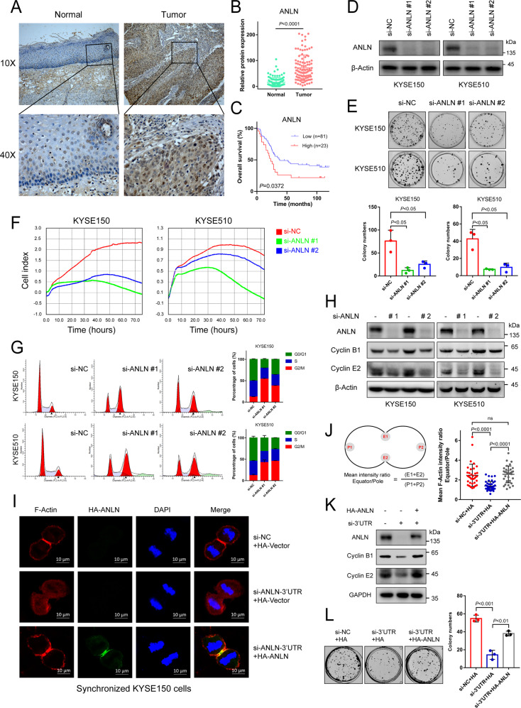
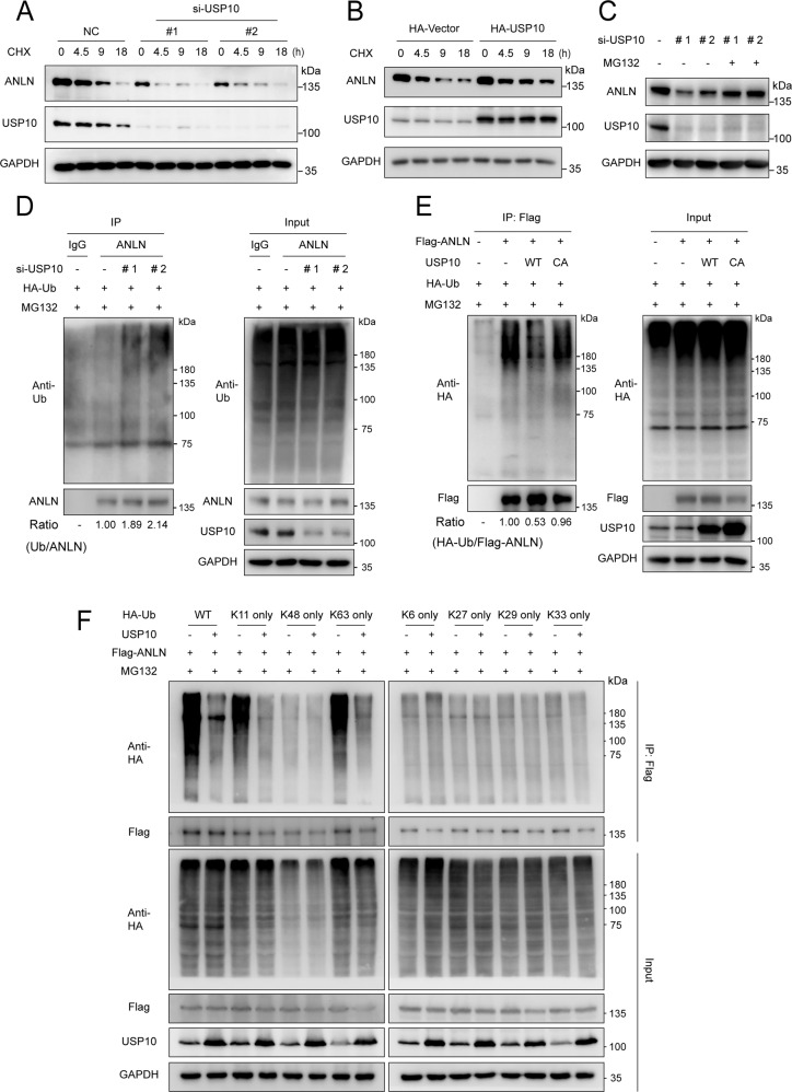
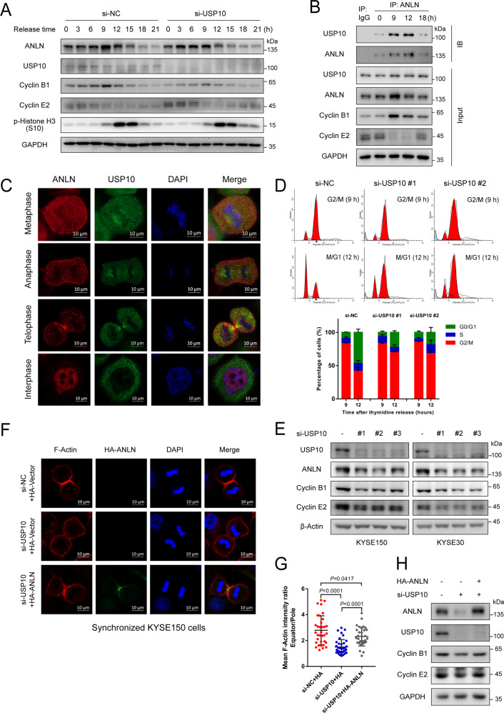
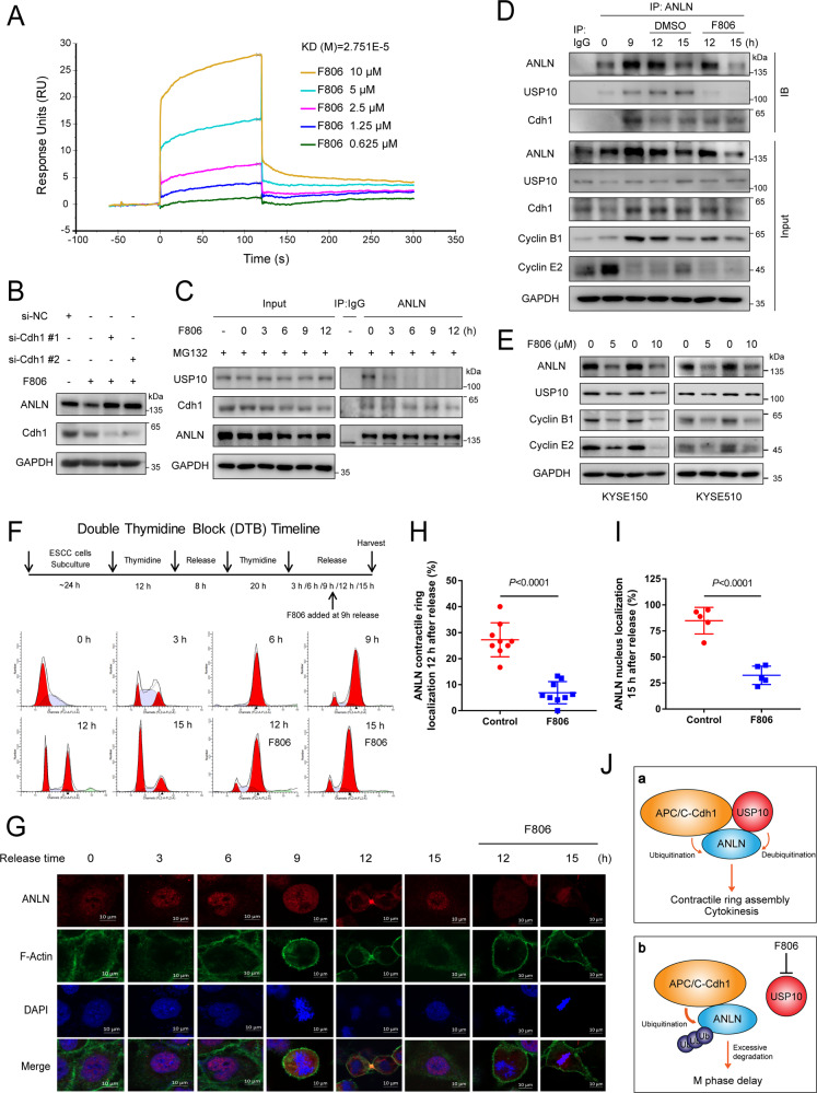
Anillin (ANLN) is a mitosis-related protein that promotes contractile ring formation and cytokinesis, but its cell cycle-dependent degradation mechanisms in cancer cells remain unclear. Here, we show that high expression of ANLN promotes cytokinesis and proliferation in esophageal squamous cell carcinoma (ESCC) cells and is associated with poor prognosis in ESCC patients. Furthermore, the findings of the study showed that the deubiquitinating enzyme USP10 interacts with ANLN and positively regulates ANLN protein levels. USP10 removes the K11- and K63-linked ubiquitin chains of ANLN through its deubiquitinase activity and prevents ANLN ubiquitin-mediated degradation. Importantly, USP10 promotes contractile ring assembly at the cytokinetic furrow as well as cytokinesis by stabilizing ANLN. Interestingly, USP10 and the E3 ubiquitin ligase APC/C co-activator Cdh1 formed a functional complex with ANLN in a non-competitive manner to balance ANLN protein levels. In addition, the macrolide compound FW-04-806 (F806), a natural compound with potential for treating ESCC, inhibited the mitosis of ESCC cells by targeting USP10 and promoting ANLN degradation. F806 selectively targeted USP10 and inhibited its catalytic activity but did not affect the binding of Cdh1 to ANLN and alters the balance of the USP10-Cdh1-ANLN complex. Additionally, USP10 expression was positively correlated with ANLN level and poor prognosis of ESCC patients. Overall, targeting the USP10-ANLN axis can effectively inhibit ESCC cell-cycle progression.


Conflict of interest statement

The authors declare no competing interests.

Figures

Fig. 1
Fig. 1. Highly expressed ANLN participates in ESCC malignant progression by promoting cytokinesis and proliferation.
A Representative image of ANLN immunohistochemical staining in ESCC and normal tissues (scale 200 and 50 μm). B ANLN expression in 104 ESCC tissues and normal tissues. C Kaplan–Meier curve analysis of the correlation between ANLN protein expression and overall survival of 104 ESCC patients. D Western blot analysis of ANLN knockdown by different siRNAs in KYSE150 and KYSE510 cells. E, F Effect of ANLN on ESCC cell proliferation was determined by colony formation (E) and an xCELLigence Real-Time Cell Analyzer (RTCA) system (F). G Cells after ANLN knockdown for 60 h were analyzed by flow cytometry. H KYSE150 cells were transfected with control or ANLN siRNA for 48 h. ANLN and cell-cycle proteins were detected using western blotting. I KYSE150 cells stably expressing HA-vector or HA-ANLN was transfected with an ANLN 3′UTR siRNA pool, then treated with thymidine for 24 h, and released for 12 h to obtain cytokinesis cells. Cells were fixed and detected by immunofluorescence. J Mean intensity ratio of individual cells were plotted (F-Actin fluorescence at the equatorial cortex: pole). P < 0.0001 by Student’s t-test. Left: diagram showing locations of regions of calculation. K–L KYSE150 cells stably expressing HA-vector or HA-ANLN were transfected with an ANLN 3’UTR siRNA pool, then cyclin B1 and E2 expression was detected by western blotting, and cell proliferation was detected by colony formation. All data are representative of at least three independent experiments and the results were statistically analyzed using a t-test.
Fig. 2
Fig. 2. USP10 interacts with ANLN and positively regulates ANLN protein level.
A Flow chart of an in vivo screen to identify candidates that interact with ANLN in KYSE150 cells. B Potential ANLN-interacting DUBs were identified by mass spectrometry analysis. C The indicated plasmids were transfected into HEK293T cells for 48 h, and then harvested with EBC buffer for co-immunoprecipitation. D-E Interaction of endogenous ANLN and USP10 in KYSE150 and KYSE30 cells was detected by co-immunoprecipitation. F, G Schematic illustration of USP10 structure (F). The indicated plasmids were transfected into HEK293T cells for 48 h, then cells were lysed and subjected to immunoprecipitation with HA magnetic beads (Thermo Fisher Scientific, 88837) (G). H, I Schematic illustration of ANLN structure (H). The indicated plasmids were transfected into HEK293T cells for 48 h, then cells were lysed and subjected to immunoprecipitation with HA magnetic beads (I). J The interaction between recombinant GST-USP10 and His-ANLN mutants was examined using an in vitro GST pull down assay. K, L ESCC cells were treated with different concentrations of spautin-1 (MedChemExpress, HY-12990) or HBX19818 (MedChemExpress, HY-17540) for 24 h, and then western blotting was used to detect the protein levels of ANLN and USP10. M KYSE150 and KYSE510 cells were transfected with siRNAs for 48 h. The indicated antibodies were used for the western blotting. N KYSE150 cells stably expressing HA-vector, HA-USP10 or HA-USP10-CA were transfected with USP10 3′ UTR siRNA, and the indicated antibodies were used for the western blotting. All data are representative of at least three independent experiments.
Fig. 3
Fig. 3. USP10 removes K11- and K63-linked ubiquitin chains of ANLN through its deubiquitinase activity.
A KYSE150 cells were transfected with siRNAs for 24 h and then treated with cycloheximide (CHX) (10 μg/ml) for different times. ANLN protein level was detected by western blotting. B KYSE150 cells stably expressing HA-vector or HA-USP10 were treated with CHX (10 μg/ml) for different times. ANLN protein level was detected by western blotting. C KYSE150 cells transfected with siRNAs were treated with MG132 (20 μM) for 10 h before harvesting, and ANLN protein levels were detected by western blotting. D KYSE150 cells transfected with siRNAs and HA-Ub were treated with MG132 (20 μM) for 10 h before harvesting, and the ubiquitination level of endogenous ANLN was measured using an ubiquitination assay. E HEK293T cells were co-transfected with Flag-ANLN, HA-Ub, USP10-WT, or CA, and treated with MG132 (20 μM) for 8 h before harvesting. Ubiquitination assays were used to detect the ubiquitination level of Flag-ANLN. F HEK293T cells were transfected with the indicated plasmids and treated with MG132 (20 μM) for 8 h before harvesting. An ubiquitination assay was used to detect the ubiquitination level of Flag-ANLN. All data are representative of at least three independent experiments.
Fig. 4
Fig. 4. USP10 promotes contractile ring localization and cytokinesis by stabilizing ANLN protein.
A KYSE150 cells transfected with siRNAs were synchronized to different time periods by thymidine. ANLN and USP10 levels were detected by western blotting. Cyclin B1, E2, and phospho-histone H3 (S10) were used as markers for the different cell-cycle phases. B KYSE150 cells were subjected to DTB and released for different time periods. Interaction between ANLN and USP10 was detected by immunoprecipitation. Cyclin B1 and E2 were used as markers for the different cell-cycle phases. C The localization of ANLN and USP10 was detected by immunofluorescence in synchronized KYSE150 cells. D KYSE150 cells transfected with siRNAs were synchronized in G2/M-phase, released for 3 h, and analyzed by flow cytometry. E KYSE150 and KYSE30 cells were transfected with siRNAs for 48 h. The indicated antibodies were used for western blotting. F KYSE150 cells stably expressing HA-vector or HA-ANLN was transfected with a USP10 siRNA pool, then treated with thymidine for 24 h, and released for 12 h to obtain cytokinesis cells. Cells were fixed and detected by immunofluorescence. G Mean intensity ratio of individual cells were plotted (F-Actin fluorescence at the equatorial cortex: pole). P < 0.0001 by Student’s t-test. H KYSE150 cells stably expressing HA-vector or HA-ANLN were transfected with a USP10 siRNA pool. The indicated antibodies were used for western blotting. All data are representative of at least three independent experiments and the results were statistically analyzed using a t-test.
Fig. 5
Fig. 5. USP10 and Cdh1 form a complex with ANLN in a non-competitive manner to balance ANLN levels.
A–C The indicated plasmids were transfected into HEK293T cells for 48 h, then cells were harvested with EBC buffer and co-immunoprecipitated using HA or Flag magnetic beads. D The interaction between the recombinant GST-Cdh1 and His-ANLN or His-USP10 was examined using an in vitro GST pull down assay. E–G KYSE150 cells were transfected with siRNAs for 40 h, and then treated with MG132 (20 μM) for 8 h. Immunoprecipitation was used to detect the interaction between endogenous USP10, Cdh1, and ANLN. H HEK293T cells were transfected with the indicated plasmids and treated with MG132 (20 μM) for 8 h before harvesting. Ubiquitination assay was used to detect the ubiquitination level of Flag-ANLN. I KYSE150 cells were transfected with siRNAs for 48 h. The indicated antibodies were used for the western blotting. J KYSE150 cells were treated with DTB and released for different time periods. Interaction between endogenous ANLN and USP10 or Cdh1 was detected by immunoprecipitation. Cyclin B1, E2, and phospho-histone H3 (S10) were used as markers for the different cell-cycle phases. K KYSE150 cells transfected with siRNAs were synchronized and released for different time periods. ANLN levels were detected by western blotting. Cyclin B1 and phospho-histone H3 (S10) were used as markers for mitosis. L–N KYSE150 cells were transfected with siRNAs for 48 h. The indicated antibodies were used for the western blotting. All data are representative of at least three independent experiments.
Fig. 6
Fig. 6. FIM-04-806 induces ANLN degradation by inhibiting USP10 deubiquitinase activity.
A Effect of F806 on ANLN expression was evaluated in xenografts (KYSE510 cells) from tumor-bearing mice. After F806 (4 mg/kg) treatment for 21 days, the level of ANLN protein in xenografts was detected by immunohistochemistry. B Cells were treated with F806 (10 μM) for 15 h and then co-treated with MG132 (10 μM) for 6 h, ANLN levels were detected by western blotting. C Cells were co-treated with cycloheximide (CHX) (10 μg/ml) and F806 (10 μM), and ANLN levels were examined by western blotting. D KYSE150 cells transfected with HA-Ub were treated with F806 (10 μM) for different times and then treated with MG132 (20 μM) for 8 h before harvest. Ubiquitination assays were performed to examine the ubiquitination level of ANLN. E KYSE150 cells transfected with Flag-vector or Flag-USP10 were treated with F806 (10 μM), and ANLN levels were detected by western blotting. F KYSE150 cells transfected with HA-USP10 or HA-USP10-CA were treated with F806 (10 μM), and ANLN levels were detected by western blotting. G KYSE150 cells were transfected with USP10 or control siRNA for 24 h and then treated with F806 (10 μM) for 16 h, and ANLN levels were detected by western blotting. H HEK293T cells were transfected with the indicated plasmids for 24 h, then treated with MG132 (10 μM) with or without F806 (10 μM) for 6 h. Subsequently, cells were harvested for ubiquitination assay. I KYSE150 cells were treated with F806 (10 μM) for 24 h. Cell lysates were subsequently labeled with HA-Ub-VS for 30 min. The indicated antibodies were used for the western blotting. J Purified USP10 protein was incubated with F806 at 25 °C for 2 h in vitro, and then labeled with HA-Ub-VS for 30 min. The indicated antibodies were used for the western blotting. K, L USP10 purified from prokaryotic cells and different concentrations of F806 were mixed at 25 °C for 2 h. Ub-AMC was then added to each well and further incubated at 37 °C for 1 h. Ub-AMC hydrolysis was measured in real time (K), and the biochemical IC50 of F806 was measured using GraphPad Prism 7 (L). All data are representative of at least three independent experiments and the results were statistically analyzed using a t-test.
Fig. 7
Fig. 7. FIM-04-806 alters the balance of the USP10-ANLN-Cdh1 complex by targeting USP10 and inhibits the M/G1 transition of ESCC cells.
A The binding between F806 and USP10 was analyzed by SPR. His-USP10 protein was immobilized on an activated CM5 sensor chip, F806 solution was then flowed across the chip. B KYSE150 cells were transfected with Cdh1 or control siRNA for 24 h and then treated with F806 (10 μM) for 12 h, and ANLN level was detected by western blotting. C KYSE150 cells were treated with F806 (10 μM) for different times and MG132 (20 μM) for 8 h before harvest. The interaction between ANLN and Cdh1 or USP10 was detected by immunoprecipitation. D KYSE150 cells were synchronized in M phase and released for different time periods with DMSO or F806 (10 μM). The interaction between ANLN and USP10 or Cdh1 was detected by immunoprecipitation. E Cells were treated with different concentrations of F806 for 16 h. The indicated antibodies were used for the western blotting. F, G Experimental protocol for DTB. KYSE150 cells were synchronized by DTB and released for different time periods. F806 (10 μM) was added at 9 h after release. The samples were analyzed by flow cytometry (F) and immunofluorescence (G). H Cells 9 h after DTB release were treated with or without F806 (10 μM) for 3 h. The contractile ring localization of ANLN was also determined. Nine fields were counted for each group. I Cells after 9 h of release were treated with or without F806 (10 μM) for 6 h. The nuclear localization of ANLN was determined. Five fields were counted in each group. All data are representative of at least three independent experiments and the results were statistically analyzed using a t-test. J Model: USP10 and Cdh1 form a functional complex with ANLN in a non-competitive manner to regulate ANLN abundance. The steady state of ANLN levels promotes contractile ring assembly and cytokinesis. Moreover, F806 selectively targets USP10 to alter the ubiquitination-deubiquitination balance of ANLN. Reduction of ANLN leads to a slowdown in the turnover of contractile ring components and a delay in M phase.
Fig. 8
Fig. 8. Expression of USP10 is positively correlated with ANLN level and poor prognosis of ESCC patients.
A Representative images of USP10 immunohistochemical staining of ESCC and normal tissues (scale 200 and 50 μm). B Expression analysis of USP10 in 104 ESCC and normal tissue samples. C USP10 protein levels in 124 ESCC and normal tissues were analyzed based on the IPX0002501000 dataset from the iProX database. D, E The mRNA levels of USP10 in tumor and adjacent normal tissues of ESCC patients were analyzed based on the GeneChip data of GSE53625 from the GEO database (D). The mRNA levels of USP10 in ESCC tumors and adjacent normal tissues were analyzed based on the SRP064894 dataset in the SRA database (E). F–I The relation between USP10 and ANLN was determined using Pearson’s correlation analysis. J–L Kaplan–Meier curve analysis of the correlation between USP10 protein expression and overall survival in ESCC patients.

References

    1. Batty P, Gerlich DW. Mitotic Chromosome Mechanics: How Cells Segregate Their Genome. Trends Cell Biol. 2019;29:717–26. doi: 10.1016/j.tcb.2019.05.007. - DOI - PubMed
    1. Perez de Castro I, de Carcer G, Montoya G, Malumbres M. Emerging cancer therapeutic opportunities by inhibiting mitotic kinases. Curr Opin Pharm. 2008;8:375–83. doi: 10.1016/j.coph.2008.06.013. - DOI - PubMed
    1. Henriques AC, Ribeiro D, Pedrosa J, Sarmento B, Silva PMA, Bousbaa H. Mitosis inhibitors in anticancer therapy: When blocking the exit becomes a solution. Cancer Lett. 2019;440-441:64–81. doi: 10.1016/j.canlet.2018.10.005. - DOI - PubMed
    1. Yan VC, Butterfield HE, Poral AH, Yan MJ, Yang KL, Pham CD, et al. Why Great Mitotic Inhibitors Make Poor Cancer Drugs. Trends Cancer. 2020;6:924–41. doi: 10.1016/j.trecan.2020.05.010. - DOI - PMC - PubMed
    1. Sivakumar S, Gorbsky GJ. Spatiotemporal regulation of the anaphase-promoting complex in mitosis. Nat Rev Mol Cell Biol. 2015;16:82–94. doi: 10.1038/nrm3934. - DOI - PMC - PubMed

Publication types

MeSH terms