

Evaluation of reverse transcriptase-polymerase spiral reaction assay for rapid and sensitive detection of severe acute respiratory syndrome coronavirus 2
- PMID: 36528050
- PMCID: PMC9750508
- DOI: 10.1016/j.cca.2022.12.009
Evaluation of reverse transcriptase-polymerase spiral reaction assay for rapid and sensitive detection of severe acute respiratory syndrome coronavirus 2
Abstract
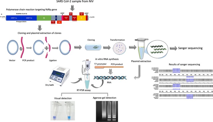
Background and aim: Existing real-time reverse transcriptase PCR (RT-qPCR) has certain limitations for the point-of-care detection of severe acute respiratory syndrome coronavirus 2 (SARS-CoV-2) since it requires sophisticated instruments, reagents and skilled laboratory personnel. In this study, we evaluated an assay termed the reverse transcriptase-polymerase spiral reaction (RT-PSR) for rapid and visual detection of SARS-CoV-2.
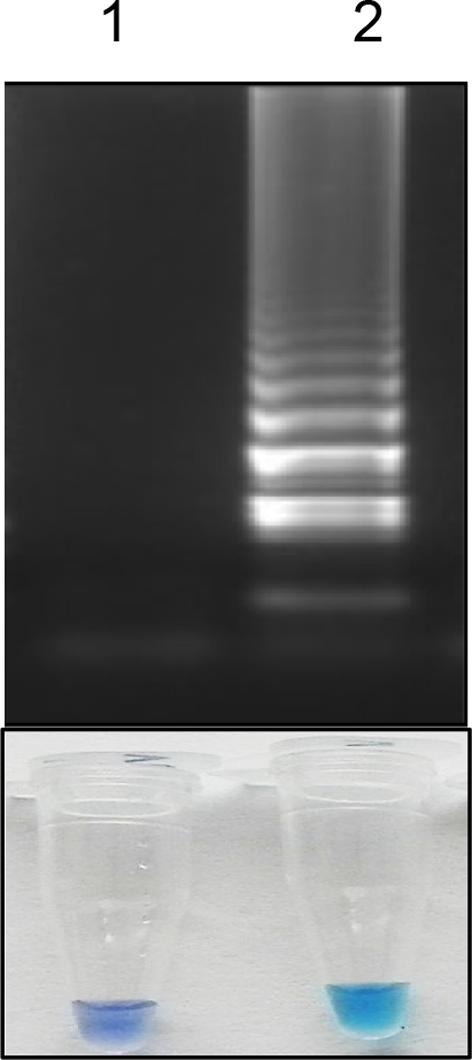
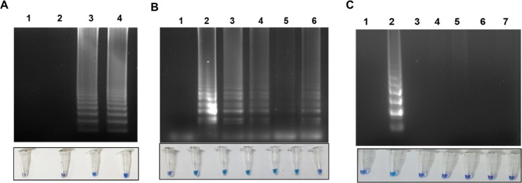
Methods: The RT-PSR assay was optimized using RdRp gene and evaluated for the detection of SARS-CoV-2. The time of 60min and a temperature of 63°C was optimized for targeting the RNA-dependent RNA polymerase gene of SARS-CoV-2. The sensitivity of the assay was evaluated by diluting the in-vitro transcribed RNA, which amplifies as low as ten copies.
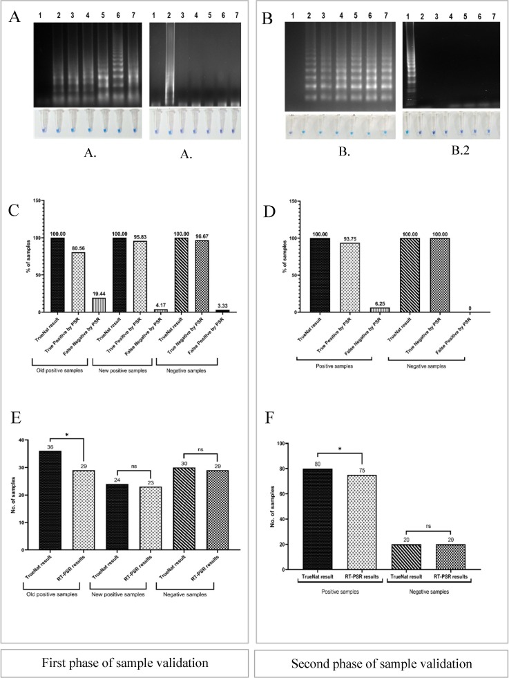
Results: The specific primers designed for this assay showed 100% specificity and did not react when tested with other lung infection-causing viruses and bacteria. The optimized assay was validated with 190 clinical samples in two phases, using automated RTPCR based TrueNat test, and the results were comparable.
Conclusions: The RT-PSR assay can be considered for rapid and sensitive detection of SARS-CoV-2, particularly in resource-limited settings. To our knowledge, there is as yet no RT-PSR-based kit developed for SARS-CoV-2.
Keywords: COVID-19; Coronavirus detection; RT-PSR; SARS-CoV-2; TrueNat test.
Copyright © 2022 Elsevier B.V. All rights reserved.
Conflict of interest statement
Declaration of Competing Interest The authors declare that they have no known competing financial interests or personal relationships that could have appeared to influence the work reported in this paper.
Figures
Similar articles
-
Comparison of a novel antigen detection test with reverse transcription polymerase chain reaction assay for laboratory diagnosis of SARS-CoV-2 infection.Infection. 2023 Feb;51(1):91-96. doi: 10.1007/s15010-022-01832-9. Epub 2022 May 5. Infection. 2023. PMID: 35513690 Free PMC article.
-
Comparison of Different Reverse Transcriptase-Polymerase Chain Reaction-Based Methods for Wastewater Surveillance of SARS-CoV-2: Exploratory Study.JMIR Public Health Surveill. 2024 Aug 19;10:e53175. doi: 10.2196/53175. JMIR Public Health Surveill. 2024. PMID: 39158943 Free PMC article.
-
Development and Clinical Application of a Rapid and Sensitive Loop-Mediated Isothermal Amplification Test for SARS-CoV-2 Infection.mSphere. 2020 Aug 26;5(4):e00808-20. doi: 10.1128/mSphere.00808-20. mSphere. 2020. PMID: 32848011 Free PMC article.
-
Combined Diagnosis of SARS-CoV-2: Rapid Antigen Detection as an Adjunct to Nucleic Acid Detection.Lab Med. 2023 Mar 7;54(2):e37-e43. doi: 10.1093/labmed/lmac089. Lab Med. 2023. PMID: 35895307 Free PMC article. Review.
-
Variability in RT-qPCR assay parameters indicates unreliable SARS-CoV-2 RNA quantification for wastewater surveillance.Water Res. 2021 Sep 15;203:117516. doi: 10.1016/j.watres.2021.117516. Epub 2021 Aug 5. Water Res. 2021. PMID: 34412018 Free PMC article. Review.
Cited by
-
Development of isothermal nucleic acid amplification technologies for rapid detection of Porcine Enterovirus-G.PLoS One. 2025 Jul 2;20(7):e0326700. doi: 10.1371/journal.pone.0326700. eCollection 2025. PLoS One. 2025. PMID: 40601569 Free PMC article.
-
Unveiling the therapeutic potential of cabozantinib-loaded poly D,L-lactic-co-glycolic acid and polysarcosine nanoparticles in inducing apoptosis and cytotoxicity in human HepG2 hepatocellular carcinoma cell lines and in vivo anti-tumor activity in SCID female mice.Front Oncol. 2023 Feb 15;13:1125857. doi: 10.3389/fonc.2023.1125857. eCollection 2023. Front Oncol. 2023. PMID: 36874145 Free PMC article.
References
MeSH terms
Substances
LinkOut - more resources
Full Text Sources
Medical
Molecular Biology Databases
Research Materials
Miscellaneous
